 Explore
 Validate
 Learn
Learn Western blot
Antibody data
- Antibody Data
- Antigen structure
- References [1]
- Comments [0]
- Validations
- Western blot [4]
- Immunocytochemistry [1]
- Immunohistochemistry [1]
- Other assay [2]
Submit
Validation data
Reference
Comment
Report error
- Product number
- PA5-22190 - Provider product page

- Provider
- Invitrogen Antibodies
- Product name
- Bcl-B Polyclonal Antibody
- Antibody type
- Polyclonal
- Antigen
- Synthetic peptide
- Description
- Recommended positive controls: Jurkat, Raji, NCI-H929.
- Concentration
- 1.32 mg/mL
Submitted references BCL2L10 Is Overexpressed in Melanoma Downstream of STAT3 and Promotes Cisplatin and ABT-737 Resistance.
Quezada MJ, Picco ME, Villanueva MB, Castro MV, Barbero G, Fernández NB, Illescas E, Lopez-Bergami P
Cancers 2020 Dec 30;13(1)
Cancers 2020 Dec 30;13(1)
No comments: Submit comment
Supportive validation
- Submitted by
- Invitrogen Antibodies (provider)
- Main image
- Experimental details
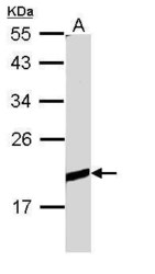
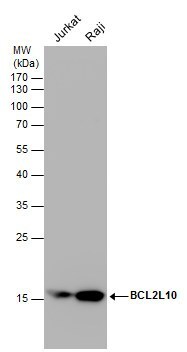
- Western blot analysis of BCL2L10 using 30 µg of Raji lysate. Samples were loaded onto a 12% SDS-PAGE gel and probed with a BCL2L10 polyclonal antibody (Product # PA5-22190) at a dilution of 1:1000.

- Submitted by
- Invitrogen Antibodies (provider)
- Main image
- Experimental details
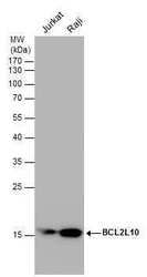
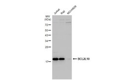
- Western Blot analysis of Bcl-B was performed by separating 30 µg of various whole cell extracts by 12% SDS-PAGE. Proteins were transferred to a membrane and probed with a Bcl-B Polyclonal Antibody (Product # PA5-22190) at a dilution of 1:1,000.
- Submitted by
- Invitrogen Antibodies (provider)
- Main image
- Experimental details
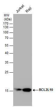
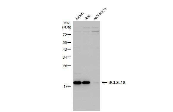
- Western Blot analysis of Bcl-B was performed by separating 30 µg of various whole cell extracts by 12% SDS-PAGE. Proteins were transferred to a membrane and probed with a Bcl-B Polyclonal Antibody (Product # PA5-22190) at a dilution of 1:1,000.
- Submitted by
- Invitrogen Antibodies (provider)
- Main image
- Experimental details
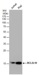
- Western Blot using Bcl-B Polyclonal Antibody (Product # PA5-22190). Various whole cell extracts (30 µg) were separated by 12% SDS-PAGE, and the membrane was blotted with Bcl-B Polyclonal Antibody (Product # PA5-22190) diluted at 1:1,000. The HRP-conjugated anti-rabbit IgG antibody was used to detect the primary antibody.
Supportive validation
- Submitted by
- Invitrogen Antibodies (provider)
- Main image
- Experimental details
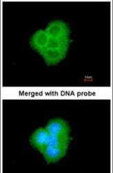
- Immunofluorescent analysis of BCL2L10 in paraformaldehyde-fixed A431 cells using a BCL2L10 polyclonal antibody (Product # PA5-22190) at a 1:200 dilution.

Supportive validation
- Submitted by
- Invitrogen Antibodies (provider)
- Main image
- Experimental details
- Immunohistochemical analysis of paraffin-embedded A549 Xenograft, using BCL2L10 (Product # PA5-22190) antibody at 1:100 dilution. Antigen Retrieval: Citrate buffer, pH 6.0, 15 min.
Supportive validation
- Submitted by
- Invitrogen Antibodies (provider)
- Main image
- Experimental details
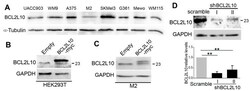
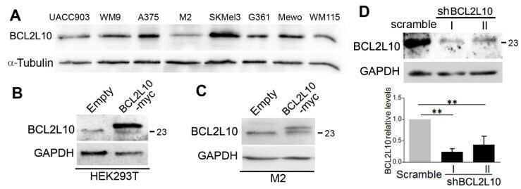
- Figure 1 BCL2L10 expression in human melanoma cell lines and validation of BCL2L10 overexpression and silencing. ( A ) Protein extracts from the indicated melanoma cell lines were probed by Western blot with a 1:1000 dilution of the BCL2L10 antibody #3869. alpha-tubulin was used as the loading control. ( B ) Validation of the BCL2L10 antibody in HEK293 transfected with BCL2L10-myc. Protein extracts from HEK293T cells transfected with BCL2L10-myc or empty plasmid were assayed by Western blot using the anti-BCL2L10 PA5-22190 antibody (1:1000 dilution). ( C ) Validation of the BCL2L10 antibody in M2 cells stably transfected with BCL2L10-myc. Protein extracts from M2-empty and M2-BCL2L10-myc cells were assayed by Western blot using the PA5-22190 antibody (1:1000 dilution). ( D ) Silencing of BCL2L10 in A375 cells. Protein extracts from A375 cells transduced with two shRNA for BCL2L10 (I and II) or a scramble shRNA were analyzed for BCL2L10 expression by Western blot using the PA5-22190 antibody (1:1000 dilution). Bar graphs show the mean +- SD (from three independent experiments) of BCL2L10 levels normalized to the loading control and expressed as the fold change relative to scramble cells. The statistical analysis is described in Methods. **: p < 0.01, n = 3. alpha-tubulin was used as a loading control in panel A and GAPDH in panels B-D. The blots displayed in all panels are representative of three independent experiments. The 23 kDa marker is indicated in panels ( B - D ). Whol
- Submitted by
- Invitrogen Antibodies (provider)
- Main image
- Experimental details
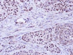
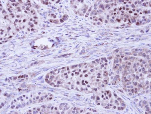
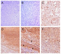
- Figure 2 BCL2L10 is highly expressed in melanoma tumor samples. Representative images of immunohistochemistry staining for BCL2L10 using the PA5-22190 antibody (1:300 dilution) with hematoxylin counterstaining. ( A , B ) Representative images from a sample that scored negative for BCL2L10 staining ( A , 100x magnification, B , 200x magnification). ( C ) Sample showing predominantly nuclear BCL2L10 staining (200x magnification). ( D , E ) Representative images from a sample showing nucleocytoplasmic BCL2L10 staining ( D , 100x magnification, E , 200x magnification). BCL2L10 staining is not observed in the adjacent stroma (at the bottom on panel D) and in the septal stroma (arrows in panel E). ( F ) Sample showing predominantly cytoplasmic BCL2L10 staining (200x magnification).
