 Explore
 Validate
 Learn
Learn Western blot
 Immunocytochemistry
Antibody data
- Antibody Data
- Antigen structure
- References [1]
- Comments [0]
- Validations
- Western blot [1]
- Immunocytochemistry [1]
- Immunohistochemistry [6]
Submit
Validation data
Reference
Comment
Report error
- Product number
- HPA000690 - Provider product page

- Provider
- Atlas Antibodies
- Proper citation
- Atlas Antibodies Cat#HPA000690, RRID:AB_1079933
- Product name
- Anti-SF3A1
- Antibody type
- Polyclonal
- Reactivity
- Human, Mouse, Rat
- Host
- Rabbit
- Conjugate
- Unconjugated
- Antigen sequence
TEDSLMPEEEFLRRNKGPVSIKVQVPNMQDKTEWK
LNGQVLVFTLPLTDQVSVIKVKIHEATGMPAGKQK
LQYEGIFIKDSNSLAYYNMANGAVIHLALKESGGR
KK- Isotype
- IgG
- Vial size
- 100 µl
- Storage
- Store at +4°C for short term storage. Long time storage is recommended at -20°C.
Submitted references The nuclear splicing factor RNA binding motif 5 promotes caspase activation in human neuronal cells, and increases after traumatic brain injury in mice.
Jackson TC, Du L, Janesko-Feldman K, Vagni VA, Dezfulian C, Poloyac SM, Jackson EK, Clark RS, Kochanek PM
Journal of cerebral blood flow and metabolism : official journal of the International Society of Cerebral Blood Flow and Metabolism 2015 Mar 31;35(4):655-66
Journal of cerebral blood flow and metabolism : official journal of the International Society of Cerebral Blood Flow and Metabolism 2015 Mar 31;35(4):655-66
No comments: Submit comment
Enhanced validation
- Submitted by
- Atlas Antibodies (provider)
- Enhanced method
- Genetic validation
- Main image

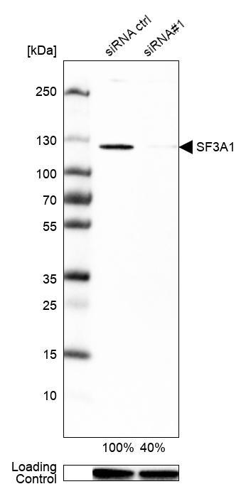
- Experimental details
- Western blot analysis in U-251MG cells transfected with control siRNA, target specific siRNA probe #1, using Anti-SF3A1 antibody. Remaining relative intensity is presented. Loading control: Anti-GAPDH.
Supportive validation
- Submitted by
- Atlas Antibodies (provider)
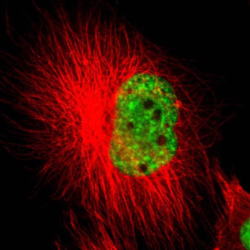
- Main image
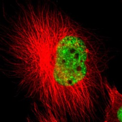
- Experimental details
- Immunofluorescent staining of human cell line U-251 MG shows localization to nuclear speckles.
- Sample type
- HUMAN
Enhanced validation
Supportive validation
- Submitted by
- Atlas Antibodies (provider)
- Enhanced method
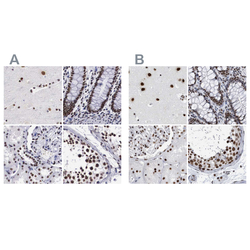
- Independent antibody validation
- Main image
- Experimental details
- Immunohistochemical staining of human cerebral cortex, colon, kidney and testis using Anti-SF3A1 antibody HPA000690 (A) shows similar protein distribution across tissues to independent antibody HPA030083 (B).

Supportive validation
- Submitted by
- Atlas Antibodies (provider)
- Main image
- Experimental details
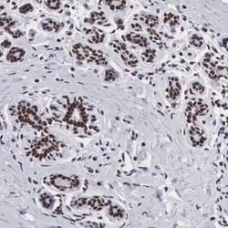
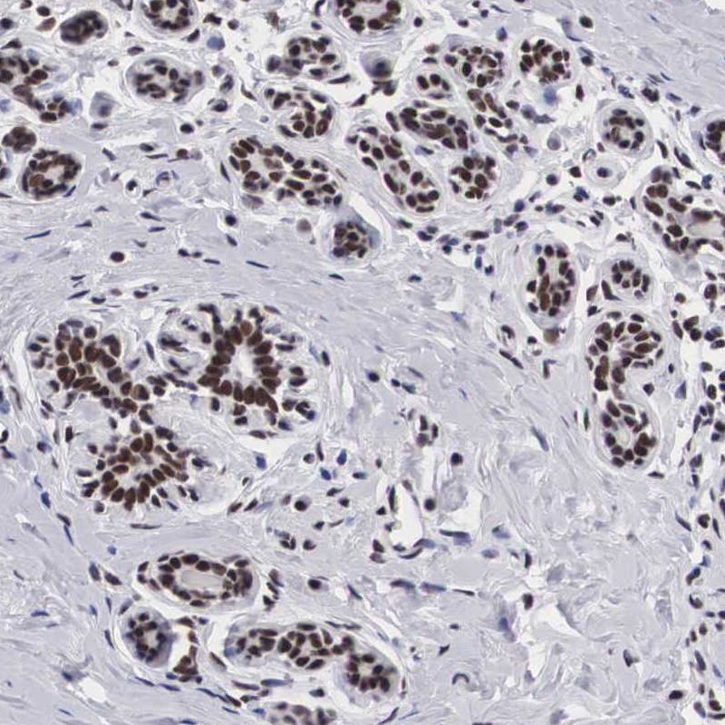
- Immunohistochemical staining of human breast shows strong nuclear positivity in glandular cells.
- Submitted by
- Atlas Antibodies (provider)
- Main image

- Experimental details
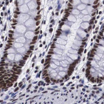
- Immunohistochemical staining of human colon using Anti-SF3A1 antibody HPA000690.
- Sample type
- HUMAN
- Submitted by
- Atlas Antibodies (provider)
- Main image

- Experimental details
- Immunohistochemical staining of human kidney using Anti-SF3A1 antibody HPA000690.
- Sample type
- HUMAN

- Submitted by
- Atlas Antibodies (provider)
- Main image
- Experimental details
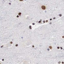
- Immunohistochemical staining of human cerebral cortex using Anti-SF3A1 antibody HPA000690.
- Sample type
- HUMAN

- Submitted by
- Atlas Antibodies (provider)
- Main image

- Experimental details
- Immunohistochemical staining of human testis using Anti-SF3A1 antibody HPA000690.
- Sample type
- HUMAN
