Explore
 Validate
 Learn
Learn Western blot
Antibody data
- Antibody Data
- Antigen structure
- References [0]
- Comments [0]
- Validations
- Western blot [5]
- Immunohistochemistry [2]
Submit
Validation data
Reference
Comment
Report error
- Product number
- PA5-18203 - Provider product page

- Provider
- Invitrogen Antibodies
- Product name
- FANCG Polyclonal Antibody
- Antibody type
- Polyclonal
- Antigen
- Synthetic peptide
- Description
- This antibody is predicted to react with canine and porcine based on sequence homology.
- Reactivity
- Human
- Host
- Goat
- Isotype
- IgG
- Vial size
- 100 µg
- Concentration
- 0.5 mg/mL
- Storage
- -20° C, Avoid Freeze/Thaw Cycles
No comments: Submit comment
Supportive validation
- Submitted by
- Invitrogen Antibodies (provider)
- Main image
- Experimental details
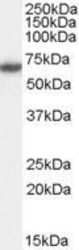
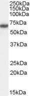
- Western Blot staining of HeLa cell lysate using Product # PA5-18203 at a concentration of 0.5 µg/mL, the primary antibody incubation was 1 hour and the detection method was chemiluminescence.

- Submitted by
- Invitrogen Antibodies (provider)
- Main image

- Experimental details
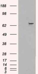
- Western blot analysis of FANCG/XRCC9 in Jurkat nuclear lysate (35µg protein in RIPA buffer). Samples were probed with the FANCG/XRCC9 antibody (Product # PA5-18203, 1µg/mL) for 1 hour. Western blot was detected by chemiluminescence.
- Submitted by
- Invitrogen Antibodies (provider)
- Main image
- Experimental details
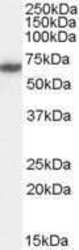
- Western Blot staining of HeLa cell lysate using Product # PA5-18203 at a concentration of 0.5 µg/mL, the primary antibody incubation was 1 hour and the detection method was chemiluminescence.
- Submitted by
- Invitrogen Antibodies (provider)
- Main image
- Experimental details
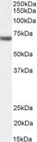
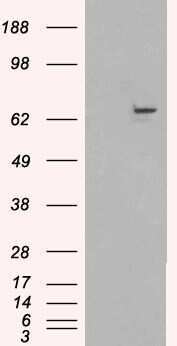
- Western blot of HEK293 overexpressing FANCG using Product # PA5-18203, mock transfection as a control in first lane.
- Submitted by
- Invitrogen Antibodies (provider)
- Main image

- Experimental details
- Western blot analysis of FANCG by a FANCG monoclonal antibody (Product # PA5-18203) at a concentration of 1 µg/mL. Jurkat nuclear lysate (35µg protein in RIPA buffer). Primary incubation was 1 hour. Detected by chemiluminescence.

Supportive validation
- Submitted by
- Invitrogen Antibodies (provider)
- Main image
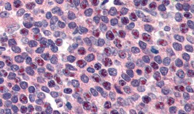
- Experimental details
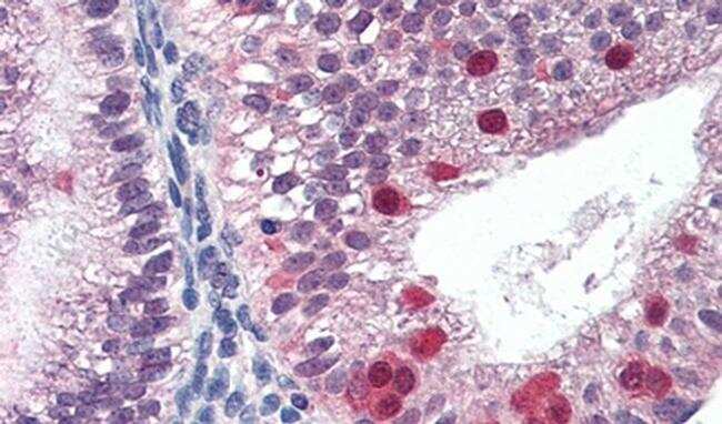
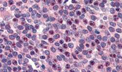
- Immunohistochemical analysis of FANCG in Human Spleen using a FANCG monoclonal antibody (Product #PA5-18203) at 3 µg/mL. The Human Spleen tissue section was paraffin embeded and detected using steamed antigen retrieval with citrate buffer pH 6, AP-staining.
- Submitted by
- Invitrogen Antibodies (provider)
- Main image

- Experimental details
- Immunohistochemical analysis of FANCG in Human Uterus using a FANCG monoclonal antibody (Product #PA5-18203) at 3 µg/mL. The Human Uterus tissue section was paraffin embeded and detected using steamed antigen retrieval with citrate buffer pH 6, AP-staining.