Explore
 Validate
 Learn
Learn Western blot
 ELISA
ELISAAntibody data
- Antibody Data
- Antigen structure
- References [0]
- Comments [0]
- Validations
- Western blot [1]
- Immunohistochemistry [1]
Submit
Validation data
Reference
Comment
Report error
- Product number
- AP09246PU-N - Provider product page

- Provider
- Acris Antibodies GmbH
- Proper citation
- Acris Antibodies GmbH Cat#AP09246PU-N, RRID:AB_2036114
- Product name
- anti XRCC9 / FANCG (1-12)
- Antibody type
- Polyclonal
- Antigen
- Synthetic peptide corresponding to amino acids 1-12 of human FANCG protein
- Reactivity
- Human
- Host
- Rabbit
- Isotype
- IgG
- Vial size
- 0.1 mg
- Concentration
- 0.93 mg/ml (by UV absorbance at 280 nm)
No comments: Submit comment
Supportive validation
- Submitted by
- Acris Antibodies GmbH (provider)
- Main image

- Experimental details
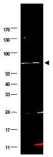
- Figure 1. Western blot using anti-FANCG antibody shows detection of a band at ~69 kDa (arrowhead) corresponding to FANCG present in a HeLa whole cell lysate. Approximately 35 μg of lysate was separated by 4-20% Tris Glycine SDS-PAGE. After blocking, the membrane was probed overnight at 4°C with the primary antibody diluted to 1:500. The membrane was washed and reacted with a 1:10,000 dilution of IRDye(TM)800 conjugated Gt-a-Rabbit IgG [H&L] for 45 min at room temperature (800 nm channel, green). Molecular weight estimation was made by comparison to prestained MW markers (indicated at left). IRDye(TM)800 fluorescence image was captured using the Odyssey® Infrared Imaging System developed by LI-COR. IRDye is a trademark of LI-COR, Inc. Other detection systems will yield similar results.
Supportive validation
- Submitted by
- Acris Antibodies GmbH (provider)
- Main image
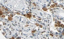
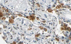
- Experimental details
- Figure 2. Immunohistochemistry. Anti-FANCG antibody shows strong nuclear and cytoplasmic staining of cells of macrophages in human lung tissue. Tissue was formalin-fixed and paraffin embedded. Brown color indicates presence of protein, blue color shows cell nuclei.Personal Communication, Kenneth Wester, www.proteinatlas.org, Uppsala, Sweden.