 Explore
 Validate
 Learn
Learn Western blot
 Immunohistochemistry
Antibody data
- Antibody Data
- Antigen structure
- References [1]
- Comments [0]
- Validations
- Western blot [5]
- Other assay [1]
Submit
Validation data
Reference
Comment
Report error
- Product number
- PA1-9091 - Provider product page

- Provider
- Invitrogen Antibodies
- Product name
- RAC2 Polyclonal Antibody
- Antibody type
- Polyclonal
- Antigen
- Synthetic peptide
- Description
- This antibody is predicted to react with mouse and rat based on sequence homology. This antibody is tested in Peptide ELISA: antibody detection limit dilution 32,000.
- Reactivity
- Human
- Host
- Goat
- Isotype
- IgG
- Vial size
- 100 µg
- Concentration
- 0.5 mg/mL
- Storage
- -20° C, Avoid Freeze/Thaw Cycles
Submitted references Time-dependent Gene Profiling Indicates the Presence of Different Phases for Ischemia/Reperfusion Injury in Retina.
Andreeva K, Zhang M, Fan W, Li X, Chen Y, Rebolledo-Mendez JD, Cooper NG
Ophthalmology and eye diseases 2014;6:43-54
Ophthalmology and eye diseases 2014;6:43-54
No comments: Submit comment
Supportive validation
- Submitted by
- Invitrogen Antibodies (provider)
- Main image

- Experimental details
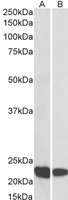
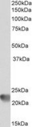
- Western blot analysis of RAC2 in Human Thymus (A) and Tonsil (B) lysate (35µg protein in RIPA buffer). Samples were probed with the RAC2 antibody (Product # PA1-9091, 0.1µg/mL) for 1 hour. Western blot was detected by chemiluminescence.

- Submitted by
- Invitrogen Antibodies (provider)
- Main image

- Experimental details
- Western blot analysis of RAC2 in Human Thymus (A) and Tonsil (B) lysate (35µg protein in RIPA buffer). Samples were probed with the RAC2 antibody (Product # PA1-9091, 0.1µg/mL) for 1 hour. Western blot was detected by chemiluminescence.
- Submitted by
- Invitrogen Antibodies (provider)
- Main image
- Experimental details
- Western blot analysis of RAC2 in Human Thymus (A) and Tonsil (B) lysate (35µg protein in RIPA buffer). Samples were probed with the RAC2 antibody (Product # PA1-9091, 0.1µg/mL) for 1 hour. Western blot was detected by chemiluminescence.

- Submitted by
- Invitrogen Antibodies (provider)
- Main image

- Experimental details
- Western blot analysis of RAC2 in Human Thymus (A) and Tonsil (B) lysate (35µg protein in RIPA buffer). Samples were probed with the RAC2 antibody (Product # PA1-9091, 0.1µg/mL) for 1 hour. Western blot was detected by chemiluminescence.

Supportive validation
- Submitted by
- Invitrogen Antibodies (provider)
- Main image
- Experimental details
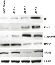
- Figure 5 Western blot analysis of protein expression in sham control and IR 0 h, 24 h and 7 d retina. C3 and Rac2 proteins reached peak accumulation at IR 7 d. Activated-caspase3 protein was increased at IR 24 h, and then gradually decreased. GNG7 protein was detected in all retinal samples, but was decreased at IR 24 h and 7 d. GRM5 protein accumulation was decreased at IR 24, then return to normal levels at IR 7 d.
