 Explore
 Validate
 Learn
Learn Western blot
Antibody data
- Antibody Data
- Antigen structure
- References [0]
- Comments [0]
- Validations
- Western blot [4]
- Immunocytochemistry [1]
- Immunohistochemistry [3]
- Other assay [1]
Submit
Validation data
Reference
Comment
Report error
- Product number
- PA5-30046 - Provider product page

- Provider
- Invitrogen Antibodies
- Product name
- GNB1 Polyclonal Antibody
- Antibody type
- Polyclonal
- Antigen
- Recombinant protein fragment
- Description
- Recommended positive controls: Jurkat, Raji, NCI-H929, Mouse brain.
- Concentration
- 1.44 mg/mL
No comments: Submit comment
Supportive validation
- Submitted by
- Invitrogen Antibodies (provider)
- Main image

- Experimental details
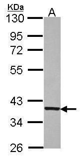
- Western blot analysis of GNB1 using 30 µg of Jurkat lysate. Samples were loaded onto a 10% SDS-PAGE gel and probed with a GNB1 polyclonal antibody (Product # PA5-30046) at a dilution of 1:5000.
- Submitted by
- Invitrogen Antibodies (provider)
- Main image
- Experimental details
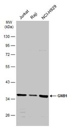
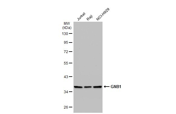
- Western Blot analysis of GNB1 was performed by separating 30 µg of various whole cell extracts by 10% SDS-PAGE. Proteins were transferred to a membrane and probed with a GNB1 Polyclonal Antibody (Product # PA5-30046) at a dilution of 1:5000 and a HRP-conjugated anti-rabbit IgG secondary antibody.

- Submitted by
- Invitrogen Antibodies (provider)
- Main image

- Experimental details
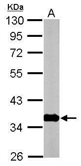
- Western Blot using GNB1 Polyclonal Antibody (Product # PA5-30046). Sample (50 µg of whole cell lysate). Lane A: mouse brain. 10% SDS PAGE. GNB1 Polyclonal Antibody (Product # PA5-30046) diluted at 1:10,000. The HRP-conjugated anti-rabbit IgG antibody was used to detect the primary antibody.
- Submitted by
- Invitrogen Antibodies (provider)
- Main image
- Experimental details
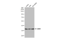
- Western Blot using GNB1 Polyclonal Antibody (Product # PA5-30046). Various whole cell extracts (30 µg) were separated by 10% SDS-PAGE, and the membrane was blotted with GNB1 Polyclonal Antibody (Product # PA5-30046) diluted at 1:5,000. The HRP-conjugated anti-rabbit IgG antibody was used to detect the primary antibody.
Supportive validation
- Submitted by
- Invitrogen Antibodies (provider)
- Main image
- Experimental details
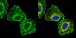
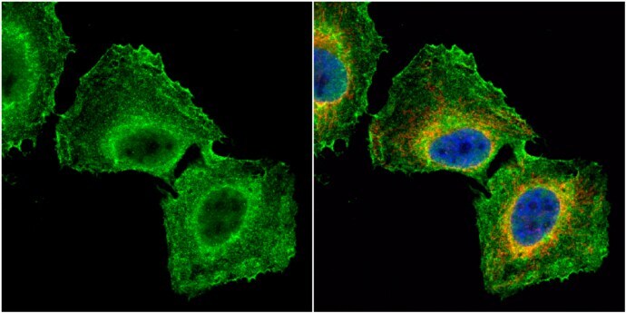
- Immunocytochemistry-Immunofluorescence analysis of GNB1 was performed in HeLa cells fixed in 4% paraformaldehyde at RT for 15 min. Green: GNB1 Polyclonal Antibody (Product # PA5-30046) diluted at 1:500. Red: FIS1, a mitochondria marker. Blue: Hoechst 33342 staining.
Supportive validation
- Submitted by
- Invitrogen Antibodies (provider)
- Main image
- Experimental details
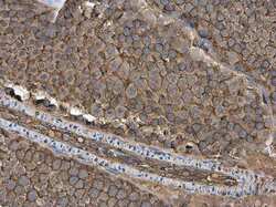
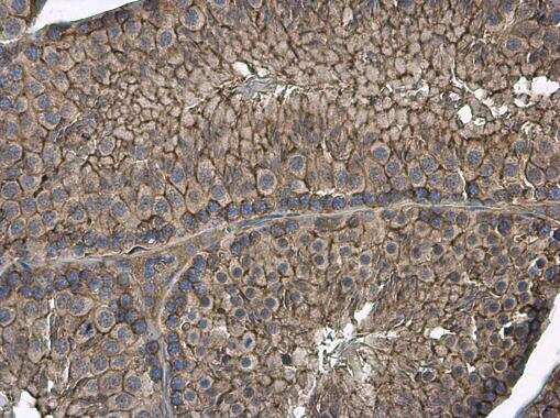
- Immunohistochemistry (Paraffin) analysis of GNB1 was performed in paraffin-embedded rat testis tissue using GNB1 Polyclonal Antibody (Product # PA5-30046) at a dilution of 1:1000.

- Submitted by
- Invitrogen Antibodies (provider)
- Main image

- Experimental details
- Immunohistochemistry (Paraffin) analysis of GNB1 was performed in paraffin-embedded mouse testis tissue using GNB1 Polyclonal Antibody (Product # PA5-30046) at a dilution of 1:1000.
- Submitted by
- Invitrogen Antibodies (provider)
- Main image
- Experimental details
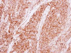
- Immunohistochemical analysis of paraffin-embedded human lung papillory adenocarcinoma, using GNB1 (Product # PA5-30046) antibody at 1:500 dilution. Antigen Retrieval: EDTA based buffer, pH 8.0, 15 min.

Supportive validation
- Submitted by
- Invitrogen Antibodies (provider)
- Main image
- Experimental details
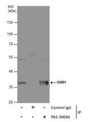
- Immunoprecipitation of GNB1 was performed in 293T whole cell extracts using 5 µg of GNB1 Polyclonal Antibody (Product # PA5-30046). Samples were transferred to a membrane and probed with GNB1 Polyclonal Antibody as a primary antibody and an HRP-conjugated anti-Rabbit IgG was used as a secondary antibody.
