 Explore
 Validate
 Learn
Learn Western blot
Antibody data
- Antibody Data
- Antigen structure
- References [1]
- Comments [0]
- Validations
- Western blot [3]
- Immunocytochemistry [1]
- Immunohistochemistry [5]
- Flow cytometry [2]
Submit
Validation data
Reference
Comment
Report error
- Product number
- TA502024 - Provider product page

- Provider
- OriGene
- Proper citation
- OriGene Cat#TA502024, RRID:AB_11124442
- Product name
- SAMHD1 mouse monoclonal antibody, clone OTI3F5 (formerly 3F5)
- Antibody type
- Monoclonal
- Description
- SAMHD1 mouse monoclonal antibody, clone OTI3F5 (formerly 3F5)
- Reactivity
- Canine
- Host
- Mouse
- Conjugate
- Unconjugated
- Epitope
- SAMHD1
- Isotype
- IgG
- Antibody clone number
- OTI3F5
- Vial size
- 100 µl
- Concentration
- 1 mg/ml
Submitted references Phosphorylation of murine SAMHD1 regulates its antiretroviral activity.
Wittmann S, Behrendt R, Eissmann K, Volkmann B, Thomas D, Ebert T, Cribier A, Benkirane M, Hornung V, Bouzas NF, Gramberg T
Retrovirology 2015 Dec 15;12:103
Retrovirology 2015 Dec 15;12:103
No comments: Submit comment
Supportive validation
- Submitted by
- OriGene (provider)
- Main image
- Experimental details
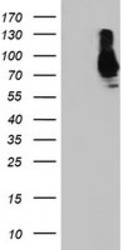
- HEK293T cells were transfected with the pCMV6-ENTRY control (Left lane) or pCMV6-ENTRY SAMHD1 (RC206013, Right lane) cDNA for 48 hrs and lysed. Equivalent amounts of cell lysates (5 ug per lane) were separated by SDS-PAGE and immunoblotted with anti-SAMHD1.
- Validation comment
- WB

- Submitted by
- OriGene (provider)
- Main image
- Experimental details
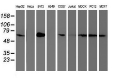
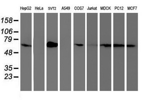
- Western blot analysis of extracts (35ug) from 9 different cell lines by using anti-SAMHD1 monoclonal antibody.
- Validation comment
- WB
- Submitted by
- OriGene (provider)
- Main image

- Experimental details
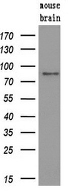
- Western blot analysis of extracts (10ug) from a mouse tissue by using anti-SAMHD1 monoclonal antibody.(1:200)
- Validation comment
- WB
Supportive validation
- Submitted by
- OriGene (provider)
- Main image

- Experimental details
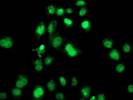
- Anti-SAMHD1 mouse monoclonal antibody (TA502024) immunofluorescent staining of COS7 cells transiently transfected by pCMV6-ENTRY SAMHD1(RC206013).
- Validation comment
- IF
Supportive validation
- Submitted by
- OriGene (provider)
- Main image

- Experimental details
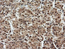
- Immunohistochemical staining of paraffin-embedded Carcinoma of Human liver tissue using anti-SAMHD1 mouse monoclonal antibody. (Heat-induced epitope retrieval by 10mM citric buffer, pH6.0, 100C for 10min, TA502024)
- Validation comment
- IHC
- Submitted by
- OriGene (provider)
- Main image
- Experimental details
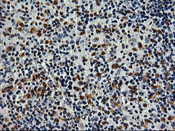
- Immunohistochemical staining of paraffin-embedded Human lymphoma tissue using anti-SAMHD1 mouse monoclonal antibody. (Heat-induced epitope retrieval by 10mM citric buffer, pH6.0, 100C for 10min, TA502024)
- Validation comment
- IHC

- Submitted by
- OriGene (provider)
- Main image
- Experimental details
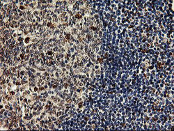
- Immunohistochemical staining of paraffin-embedded Human tonsil within the normal limits using anti-SAMHD1 mouse monoclonal antibody. (Heat-induced epitope retrieval by 10mM citric buffer, pH6.0, 100C for 10min, TA502024)
- Validation comment
- IHC

- Submitted by
- OriGene (provider)
- Main image

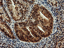
- Experimental details
- Immunohistochemical staining of paraffin-embedded Adenocarcinoma of Human endometrium tissue using anti-SAMHD1 mouse monoclonal antibody. (Heat-induced epitope retrieval by 10mM citric buffer, pH6.0, 100C for 10min, TA502024)
- Validation comment
- IHC
- Submitted by
- OriGene (provider)
- Main image
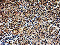
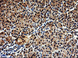
- Experimental details
- Immunohistochemical staining of paraffin-embedded Human pancreas tissue within the normal limits using anti-SAMHD1 mouse monoclonal antibody. (Heat-induced epitope retrieval by 10mM citric buffer, pH6.0, 100C for 10min, TA502024)
- Validation comment
- IHC
Supportive validation
- Submitted by
- OriGene (provider)
- Main image
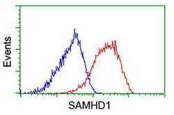
- Experimental details
- Flow cytometric Analysis of Hela cells, using anti-SAMHD1 antibody(TA502024),(Red), compared to a nonspecific negative control antibody,(Blue).
- Validation comment
- FC

- Submitted by
- OriGene (provider)
- Main image

- Experimental details
- Flow cytometric Analysis of Jurkat cells, using anti-SAMHD1 antibody(TA502024),(Red), compared to a nonspecific negative control antibody,(Blue).
- Validation comment
- FC
