 Explore
 Validate
 Learn
Learn Western blot
Antibody data
- Antibody Data
- Antigen structure
- References [3]
- Comments [0]
- Validations
- Western blot [1]
- Immunohistochemistry [1]
Submit
Validation data
Reference
Comment
Report error
- Product number
- AF5725 - Provider product page

- Provider
- Novus Biologicals
- Product name
- Sheep Polyclonal Corneodesmosin Antibody
- Antibody type
- Polyclonal
- Description
- Immunogen affinity purified. Detects human Corneodesmosin in direct ELISAs and Western blots.
- Reactivity
- Human
- Host
- Sheep
- Isotype
- IgG
- Vial size
- 100 ug
- Concentration
- LYOPH
- Storage
- Use a manual defrost freezer and avoid repeated freeze-thaw cycles. 12 months from date of receipt, -20 to -70 degreesC as supplied. 1 month, 2 to 8 degreesC under sterile conditions after reconstitution. 6 months, -20 to -70 degreesC under sterile conditions after reconstitution.
Submitted references Pharmacological modulators of autophagy activate a parallel noncanonical pathway driving unconventional LC3 lipidation.
The caspase-1 inhibitor CARD18 is specifically expressed during late differentiation of keratinocytes and its expression is lost in lichen planus.
Callus formation is associated with hyperproliferation and incomplete differentiation of keratinocytes, and increased expression of adhesion molecules.
Jacquin E, Leclerc-Mercier S, Judon C, Blanchard E, Fraitag S, Florey O
Autophagy 2017 May 4;13(5):854-867
Autophagy 2017 May 4;13(5):854-867
The caspase-1 inhibitor CARD18 is specifically expressed during late differentiation of keratinocytes and its expression is lost in lichen planus.
Qin H, Jin J, Fischer H, Mildner M, Gschwandtner M, Mlitz V, Eckhart L, Tschachler E
Journal of dermatological science 2017 Aug;87(2):176-182
Journal of dermatological science 2017 Aug;87(2):176-182
Callus formation is associated with hyperproliferation and incomplete differentiation of keratinocytes, and increased expression of adhesion molecules.
Kim SH, Kim S, Choi HI, Choi YJ, Lee YS, Sohn KC, Lee Y, Kim CD, Yoon TJ, Lee JH, Lee YH
The British journal of dermatology 2010 Sep;163(3):495-501
The British journal of dermatology 2010 Sep;163(3):495-501
No comments: Submit comment
Supportive validation
- Submitted by
- Novus Biologicals (provider)
- Main image
- Experimental details
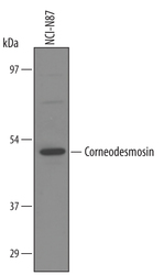
- Detection of Human Corneodesmosin by Western Blot. Western blot shows lysates of NCI-N87 human gastric carcinoma cell line. PVDF membrane was probed with 1 µg/mL of Sheep Anti-Human Corneodesmosin Antigen Affinity-purified Polyclonal Antibody (Catalog # AF5725) followed by HRP-conjugated Anti-Sheep IgG Secondary Antibody (Catalog # HAF016). A specific band was detected for Corneodesmosin at approximately 52 kDa (as indicated). This experiment was conducted under reducing conditions and using Immunoblot Buffer Group 8.

Supportive validation
- Submitted by
- Novus Biologicals (provider)
- Main image
- Experimental details
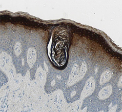
- Corneodesmosin in Human Skin. Corneodesmosin was detected in immersion fixed paraffin-embedded sections of human skin using Sheep Anti-Human Corneodesmosin Antigen Affinity-purified Polyclonal Antibody (Catalog # AF5725) at 1.7 µg/mL overnight at 4 °C. Tissue was stained using the Anti-Sheep HRP-DAB Cell & Tissue Staining Kit (brown; Catalog # CTS019) and counterstained with hematoxylin (blue). View our protocol for Chromogenic IHC Staining of Paraffin-embedded Tissue Sections.
