Explore
 Validate
 Learn
Learn Western blot
 Immunocytochemistry
Antibody data
- Antibody Data
- Antigen structure
- References [2]
- Comments [0]
- Validations
- Immunocytochemistry [1]
Submit
Validation data
Reference
Comment
Report error
- Product number
- MAB662 - Provider product page

- Provider
- R&D Systems
- Product name
- Mouse OSMR beta Antibody
- Antibody type
- Monoclonal
- Description
- Protein A or G purified from hybridoma culture supernatant. Detects mouse OSM R beta in direct ELISAs and Western blots. In Western blots, no cross-reactivity with recombinant human (rh) CLC, rhCNTF, rhOSM, recombinant mouse (rm) CT-1, rmIL-6, rmIL-11, or rmLIF is observed.
- Reactivity
- Mouse
- Host
- Rat
- Conjugate
- Unconjugated
- Antigen sequence
O70458- Isotype
- IgG
- Antibody clone number
- 118125
- Vial size
- 100 ug
- Concentration
- LYOPH
- Storage
- Use a manual defrost freezer and avoid repeated freeze-thaw cycles. 12 months from date of receipt, -20 to -70 °C as supplied. 1 month, 2 to 8 °C under sterile conditions after reconstitution. 6 months, -20 to -70 °C under sterile conditions after reconstitution.
Submitted references Oncostatin M induces dendritic cell maturation and Th1 polarization.
Characterization of the potential subpopulation of bone marrow cells involved in the repair of injured liver tissue.
Jung ID, Noh KT, Lee CM, Chun SH, Jeong SK, Park JW, Park WS, Kim HW, Yun CH, Shin YK, Park YM
Biochemical and biophysical research communications 2010 Apr 2;394(2):272-8
Biochemical and biophysical research communications 2010 Apr 2;394(2):272-8
Characterization of the potential subpopulation of bone marrow cells involved in the repair of injured liver tissue.
Khurana S, Mukhopadhyay A
Stem cells (Dayton, Ohio) 2007 Jun;25(6):1439-47
Stem cells (Dayton, Ohio) 2007 Jun;25(6):1439-47
No comments: Submit comment
Supportive validation
- Submitted by
- R&D Systems (provider)
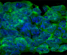
- Main image

- Experimental details
- OSM R beta in D3 Mouse Cell Line. OSM R beta was detected in immersion fixed D3 mouse embryonic stem cell line using Rat Anti-Mouse OSM R beta Monoclonal Antibody (Catalog # MAB662) at 10 µg/mL for 3 hours at room temperature. Cells were stained using the NorthernLights™ 493-conjugated Anti-Rat IgG Secondary Antibody (green; Catalog # NL015) and counterstained with DAPI (blue). Specific staining was localized to cytoplasm and plasma membrane. View our protocol for Fluorescent ICC Staining of Cells on Coverslips.