 Explore
 Validate
 Learn
Learn Western blot
Antibody data
- Antibody Data
- Antigen structure
- References [3]
- Comments [0]
- Validations
- Western blot [2]
- Immunocytochemistry [2]
- Immunohistochemistry [1]
- Other assay [1]
Submit
Validation data
Reference
Comment
Report error
- Product number
- PA5-21721 - Provider product page

- Provider
- Invitrogen Antibodies
- Product name
- ST3GAL1 Polyclonal Antibody
- Antibody type
- Polyclonal
- Antigen
- Recombinant protein fragment
- Description
- Recommended positive controls: HeLa.
- Concentration
- 0.63 mg/mL
Submitted references Sialylation of TLR2 initiates osteoclast fusion.
ST3GAL1 is a target of the SOX2-GLI1 transcriptional complex and promotes melanoma metastasis through AXL.
Towards Age-Related Anti-Inflammatory Therapy: Klotho Suppresses Activation of ER and Golgi Stress Response in Senescent Monocytes.
Dou C, Zhen G, Dan Y, Wan M, Limjunyawong N, Cao X
Bone research 2022 Mar 2;10(1):24
Bone research 2022 Mar 2;10(1):24
ST3GAL1 is a target of the SOX2-GLI1 transcriptional complex and promotes melanoma metastasis through AXL.
Pietrobono S, Anichini G, Sala C, Manetti F, Almada LL, Pepe S, Carr RM, Paradise BD, Sarkaria JN, Davila JI, Tofani L, Battisti I, Arrigoni G, Ying L, Zhang C, Li H, Meves A, Fernandez-Zapico ME, Stecca B
Nature communications 2020 Nov 17;11(1):5865
Nature communications 2020 Nov 17;11(1):5865
Towards Age-Related Anti-Inflammatory Therapy: Klotho Suppresses Activation of ER and Golgi Stress Response in Senescent Monocytes.
Mytych J, Sołek P, Będzińska A, Rusinek K, Warzybok A, Tabęcka-Łonczyńska A, Koziorowski M
Cells 2020 Jan 21;9(2)
Cells 2020 Jan 21;9(2)
No comments: Submit comment
Supportive validation
- Submitted by
- Invitrogen Antibodies (provider)
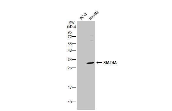
- Main image

- Experimental details
- Western blot analysis of ST3GAL1 using 30 µg of HeLa S3 lysate. Samples were loaded onto a 12% SDS-PAGE gel and probed with a ST3GAL1 polyclonal antibody (Product # PA5-21721) at a dilution of 1:500.

- Submitted by
- Invitrogen Antibodies (provider)
- Main image

- Experimental details
- Western Blot using ST3GAL1 Polyclonal Antibody (Product # PA5-21721). Various whole cell extracts (30 µg) were separated by 12% SDS-PAGE, and the membrane was blotted with ST3GAL1 Polyclonal Antibody (Product # PA5-21721) diluted at 1:500. The HRP-conjugated anti-rabbit IgG antibody was used to detect the primary antibody.
Supportive validation
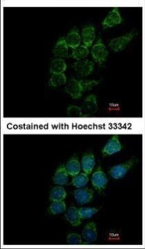
- Submitted by
- Invitrogen Antibodies (provider)
- Main image

- Experimental details
- Immunofluorescent analysis of ST3GAL1 in methanol-fixed Hep3B cells using a ST3GAL1 polyclonal antibody (Product # PA5-21721) at a 1:500 dilution.
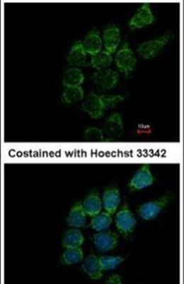
- Submitted by
- Invitrogen Antibodies (provider)
- Main image

- Experimental details
- Immunofluorescence analysis of methanol-fixed Hep3B, using SIAT4A antibody (Product # PA5-21721) at 1:500 dilution.
Supportive validation
- Submitted by
- Invitrogen Antibodies (provider)
- Main image
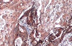
- Experimental details
- Immunohistochemistry (Paraffin) analysis of ST3GAL1 was performed in paraffin-embedded human ovarian cancer tissue using ST3GAL1 Polyclonal Antibody (Product # PA5-21721) at a dilution of 1:500. Antigen Retrieval: Citrate buffer, pH 6.0, 15 min.

Supportive validation
- Submitted by
- Invitrogen Antibodies (provider)
- Main image
- Experimental details
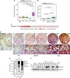
- Fig. 2 Expression of ST3GAL1 in cutaneous melanomas and association with melanoma progression. a , b Box plots illustrating expression of ST3GAL1 mRNA in skin cancers ( a ) and in nevi, primary and metastatic melanoma samples ( b ). Box-plots report median (central lines), 25th and 75th percentiles (box limits), and upper and lower whiskers represent values no further than x1.5 interquartile range (IQR). Data were obtained from the analysis of the public available microarray data sets GSE7553 and GSE46517, respectively. In GSE7553: normal skin ( n = 4), in situ melanomas (MIS) ( n = 2), primary and metastatic melanomas ( n = 54), basal cell carcinomas (BCC) ( n = 15), and squamous cell carcinomas (SCC) ( n = 11). In GSE46517: nevi ( n = 9), primary melanomas ( n = 31), metastatic melanomas ( n = 73). P value was calculated by ANOVA and Holm-Sidak's test. c Genomic profile of ST3GAL1 in melanoma patients obtained from Skin Cutaneous Melanoma data set (TCGA, Provisional) using cBioportal database ( http://www.cbioportal.org ) , . In all, 23% of melanoma samples present alterations of ST3GAL1 , including gene amplification, mRNA upregulation or somatic mutations. d Immunostaining of ST3GAL1 in human melanoma tissue microarrays, including nevi ( n = 23), primary ( n = 56), and metastatic ( n = 40) malignant melanomas. Representative images of human normal skin, nevi, primary (PM), and metastatic melanomas (MM). ST3GAL1 staining was evaluated blindly and scored as negative (no sig
