 Explore
 Validate
 Learn
Learn Western blot
 Immunocytochemistry
Antibody data
- Antibody Data
- Antigen structure
- References [2]
- Comments [0]
- Validations
- Western blot [2]
- Immunoprecipitation [1]
- Immunohistochemistry [3]
Submit
Validation data
Reference
Comment
Report error
- Product number
- NB600-1469 - Provider product page

- Provider
- Novus Biologicals
- Proper citation
- Novus Cat#NB600-1469, RRID:AB_2119820
- Product name
- Mouse Monoclonal HSP70/HSPA1A Antibody
- Antibody type
- Monoclonal
- Description
- Unpurified. Epitope mapping with a panel of HSP70 deletion mutants suggests that the epitope recognized is located between amino acids 504-617 of human HSP70, a region that has been shown to be involved in stress-induced nucleolar localization.
- Reactivity
- Human, Mouse, Rat, Chicken/Avian, Drosophila, Porcine, Simian, Yeast
- Host
- Mouse
- Isotype
- IgG
- Vial size
- 50uL
- Storage
- Store at -20C. Avoid freeze-thaw cycles.
Submitted references Acidic microenvironment plays a key role in human melanoma progression through a sustained exosome mediated transfer of clinically relevant metastatic molecules.
Blocking Hsp70 enhances the efficiency of amphotericin B treatment against resistant Aspergillus terreus strains.
Boussadia Z, Lamberti J, Mattei F, Pizzi E, Puglisi R, Zanetti C, Pasquini L, Fratini F, Fantozzi L, Felicetti F, Fecchi K, Raggi C, Sanchez M, D'Atri S, Carè A, Sargiacomo M, Parolini I
Journal of experimental & clinical cancer research : CR 2018 Oct 5;37(1):245
Journal of experimental & clinical cancer research : CR 2018 Oct 5;37(1):245
Blocking Hsp70 enhances the efficiency of amphotericin B treatment against resistant Aspergillus terreus strains.
Blatzer M, Blum G, Jukic E, Posch W, Gruber P, Nagl M, Binder U, Maurer E, Sarg B, Lindner H, Lass-Flörl C, Wilflingseder D
Antimicrobial agents and chemotherapy 2015 Jul;59(7):3778-88
Antimicrobial agents and chemotherapy 2015 Jul;59(7):3778-88
No comments: Submit comment
Supportive validation
- Submitted by
- Novus Biologicals (provider)
- Main image
- Experimental details
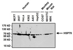
- Western Blot: HSP70/HSPA1A Antibody (3A3) [NB600-1469] - Proteins were transferred to a PVDF membrane and blocked with 5% BSA/TBST for at least 1 hour. The membrane was probed with a Hsp70 monoclonal antibody at a dilution of 1:1000 overnight at 4C on a rocking platform, washed in TBS-0.1%Tween 20, and probed with a goat anti-mouse IgG-HRP secondary antibody at a dilution of 1:20,000 for at least 1 hour.

- Submitted by
- Novus Biologicals (provider)
- Main image
- Experimental details
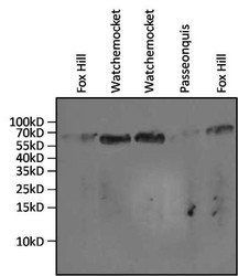
- Western Blot: HSP70/HSPA1A Antibody (3A3) [NB600-1469] - Analysis of 20ul of gill tissue lysates from the salt marsh mussel, Guekensia demissa, isolated from various coves in Rhode Island (indicated above the lanes).

Supportive validation
- Submitted by
- Novus Biologicals (provider)
- Main image
- Experimental details
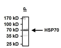
- Immunoprecipitation: HSP70/HSPA1A Antibody (3A3) [NB600-1469] - Immunoprecipitation of Heat Shock Protein 70 (Hsp70) was performed on HeLa cells. Antigen:antibody complexes were formed by incubating 500ug whole cell lysate with 2ug of Hsp70 monoclonal antibody overnight on a rocking platform at 4C. The immune complexes were captured on 50ul Protein A/G Plus Agarose at a dilution of 1:1000 overnight rotating at 4C, washed in TBST, and probed with goat anti-mouse IgG-HRP secondary antibody at a dilution of 1:20,000 for at least 1 hour.

Supportive validation
- Submitted by
- Novus Biologicals (provider)
- Main image
- Experimental details
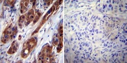
- Immunohistochemistry-Paraffin: HSP70/HSPA1A Antibody (3A3) [NB600-1469] - Both normal and cancer biopsies of deparaffinized human Breast carcinoma tissue.

- Submitted by
- Novus Biologicals (provider)
- Main image

- Experimental details
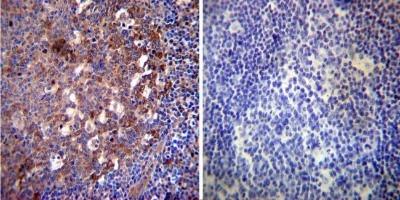
- Immunohistochemistry-Paraffin: HSP70/HSPA1A Antibody (3A3) [NB600-1469] - Both normal and cancer biopsies of deparaffinized human Tonsil tissue.
- Submitted by
- Novus Biologicals (provider)
- Main image
- Experimental details
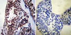
- Immunohistochemistry-Paraffin: HSP70/HSPA1A Antibody (3A3) [NB600-1469] - Both normal and cancer biopsies of deparaffinized human Testis tissue.
