 Explore
 Validate
 Learn
Learn Western blot
 Immunohistochemistry
Antibody data
- Antibody Data
- Antigen structure
- References [0]
- Comments [0]
- Validations
- Western blot [4]
- Immunohistochemistry [5]
Submit
Validation data
Reference
Comment
Report error
- Product number
- HPA037893 - Provider product page

- Provider
- Atlas Antibodies
- Proper citation
- Atlas Antibodies Cat#HPA037893, RRID:AB_10672468
- Product name
- Anti-TNIP1
- Antibody type
- Polyclonal
- Reactivity
- Human, Mouse, Rat
- Host
- Rabbit
- Conjugate
- Unconjugated
- Antigen sequence
MEGRGPYRIYDPGGSVPSGEASAAFERLVKENSRL
KEKMQGIKMLGELLEESQMEATRLRQKAEELVKDN
ELLPP- Isotype
- IgG
- Vial size
- 100 µl
- Storage
- Store at +4°C for short term storage. Long time storage is recommended at -20°C.
No comments: Submit comment
Supportive validation
Supportive validation
- Submitted by
- Atlas Antibodies (provider)
- Enhanced method
- Genetic validation
- Main image
- Experimental details
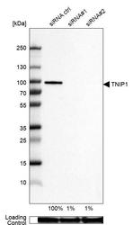
- Western blot analysis in U-87MG ATCC cells transfected with control siRNA, target specific siRNA probe #1 and #2, using Anti-TNIP1 antibody. Remaining relative intensity is presented. Loading control: Anti-GAPDH.
- Submitted by
- Atlas Antibodies (provider)
- Enhanced method
- Independent antibody validation
- Main image
- Experimental details
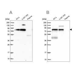
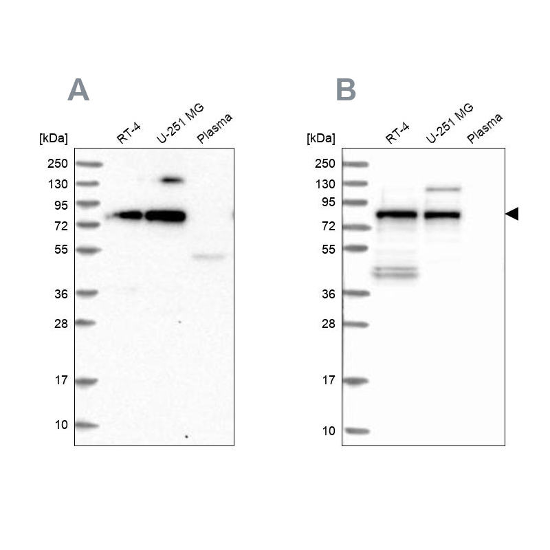
- Western blot analysis using Anti-TNIP1 antibody HPA037893 (A) shows similar pattern to independent antibody HPA037894 (B).
- Sample type
- HUMAN
Supportive validation
- Submitted by
- Atlas Antibodies (provider)
- Main image
- Experimental details
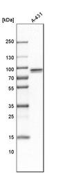
- Western blot analysis in human cell line A-431.

- Submitted by
- Atlas Antibodies (provider)
- Main image
- Experimental details
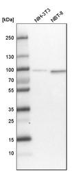
- Western blot analysis in mouse cell line NIH-3T3 and rat cell line NBT-II.

Supportive validation
- Submitted by
- Atlas Antibodies (provider)
- Main image

- Experimental details
- Immunohistochemical staining of human heart muscle shows strong cytoplasmic positivity in myocytes.
- Submitted by
- Atlas Antibodies (provider)
- Main image

- Experimental details
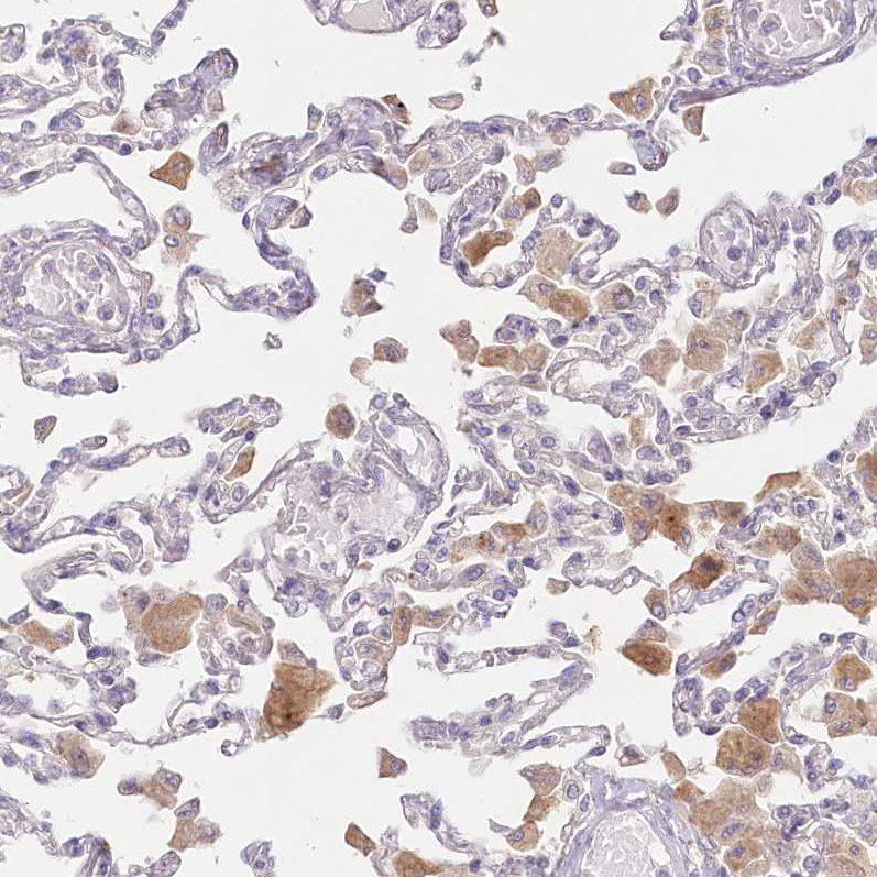
- Immunohistochemical staining of human lung shows moderate cytoplasmic positivity in macrophages.
- Sample type
- HUMAN
- Submitted by
- Atlas Antibodies (provider)
- Main image
- Experimental details
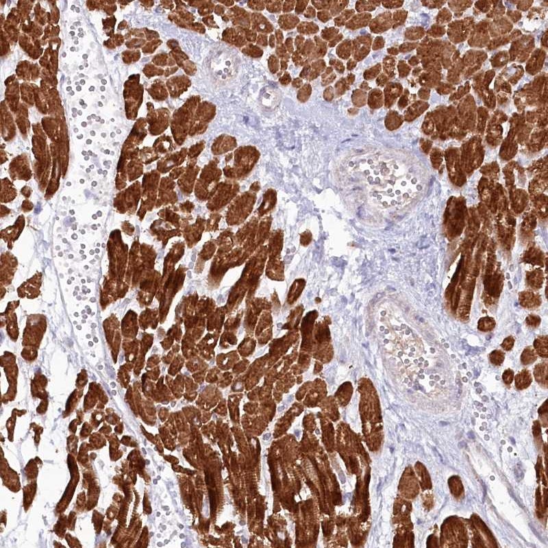
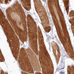
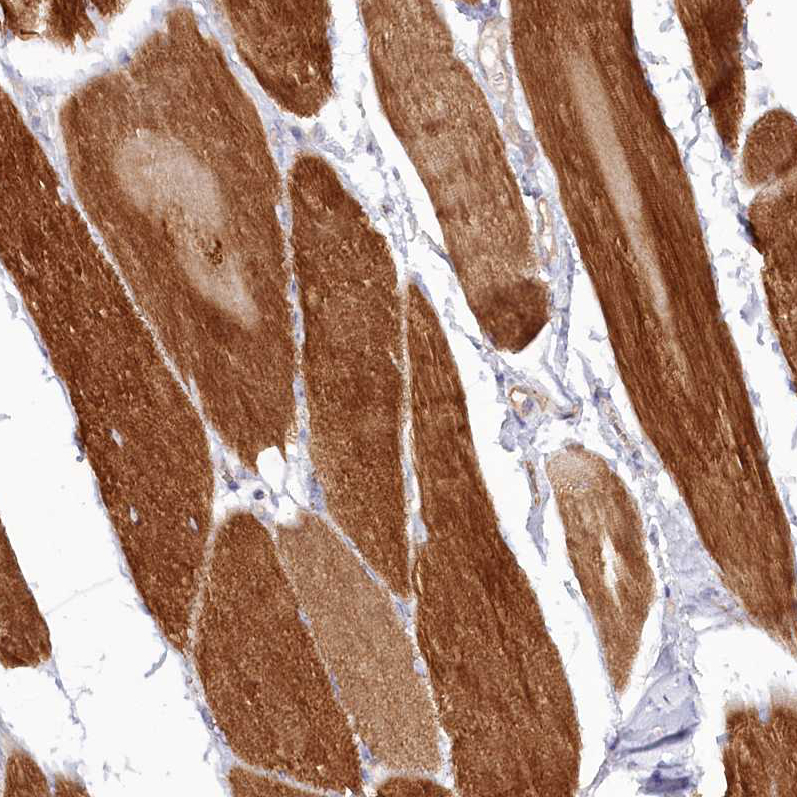
- Immunohistochemical staining of human skeletal muscle shows strong cytoplasmic positivity in myocytes.
- Sample type
- HUMAN
- Submitted by
- Atlas Antibodies (provider)
- Main image
- Experimental details
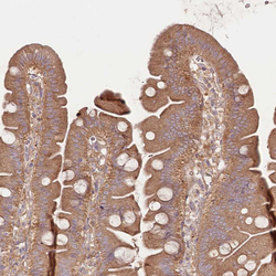
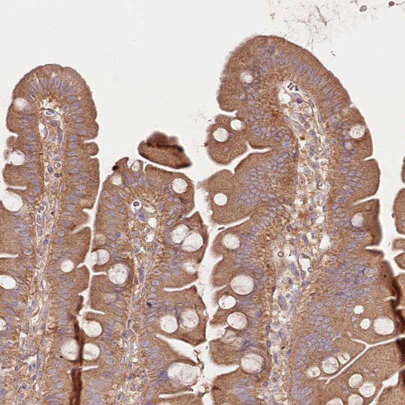
- Immunohistochemical staining of human small intestine shows moderate cytoplasmic positivity in glandular cells.
- Sample type
- HUMAN
- Submitted by
- Atlas Antibodies (provider)
- Main image
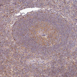
- Experimental details
- Immunohistochemical staining of human lymph node shows moderate cytoplasmic positivity in germinal center cells.
- Sample type
- HUMAN
