Explore
 Validate
 Learn
Learn Western blot
 Immunocytochemistry
 Immunoprecipitation
Antibody data
- Antibody Data
- Antigen structure
- References [2]
- Comments [0]
- Validations
- Western blot [10]
- Immunocytochemistry [2]
- Immunohistochemistry [1]
Submit
Validation data
Reference
Comment
Report error
- Product number
- GTX103005 - Provider product page

- Provider
- GeneTex
- Proper citation
- GeneTex Cat#GTX103005, RRID:AB_1950046
- Product name
- CSE1L antibody
- Antibody type
- Polyclonal
- Reactivity
- Human, Mouse, Rat
- Host
- Rabbit
Submitted references A pharmacological probe identifies cystathionine β-synthase as a new negative regulator for ferroptosis.
Ets-1 facilitates nuclear entry of NFAT proteins and their recruitment to the IL-2 promoter.
Wang L, Cai H, Hu Y, Liu F, Huang S, Zhou Y, Yu J, Xu J, Wu F
Cell death & disease 2018 Sep 26;9(10):1005
Cell death & disease 2018 Sep 26;9(10):1005
Ets-1 facilitates nuclear entry of NFAT proteins and their recruitment to the IL-2 promoter.
Tsao HW, Tai TS, Tseng W, Chang HH, Grenningloh R, Miaw SC, Ho IC
Proceedings of the National Academy of Sciences of the United States of America 2013 Sep 24;110(39):15776-81
Proceedings of the National Academy of Sciences of the United States of America 2013 Sep 24;110(39):15776-81
No comments: Submit comment
Enhanced validation
Supportive validation
- Submitted by
- GeneTex (provider)
- Enhanced method
- Genetic validation
- Main image
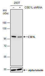
- Experimental details
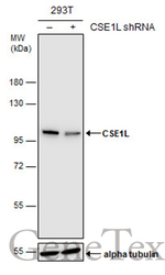
- Non-transfected (¡V) and transfected (+) 293T whole cell extracts (15 ?g) were separated by 7.5% SDS-PAGE, and the membrane was blotted with CSE1L antibody (GTX103005) diluted at 1:5000. The HRP-conjugated anti-rabbit IgG antibody (GTX213110-01) was used to detect the primary antibody.
Supportive validation
- Submitted by
- GeneTex (provider)
- Main image
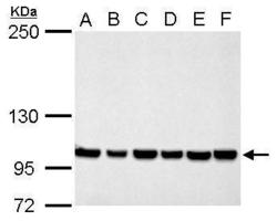
- Experimental details
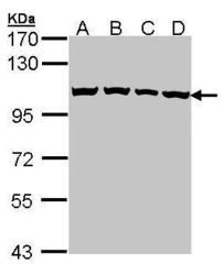
- CSE1L antibody detects CSE1L protein by Western blot analysis.A. 30 µg Neuro2A whole cell lysate/extract B. 30 µg GL261 whole cell lysate/extract C. 30 µg C8D30 whole cell lysate/extract D. 30 µg NIH-3T3 whole cell lysate/extract E. 30 µg BCL-1 whole cell lysate/extract F. 30 µg Raw264.7 whole cell lysate/extract G. 30 µg C2C12 whole cell lysate/extract 5 % SDS-PAGECSE1L antibody (GTX103005) dilution: 1:1000
- Validation comment
- WB

- Submitted by
- GeneTex (provider)
- Main image
- Experimental details
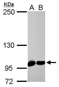
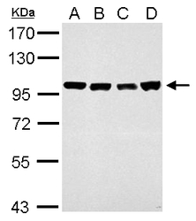
- Sample (30 ug of whole cell lysate) A: 293T B: A431 (GTX27909) C: H1299 D: Hela 7.5% SDS PAGE GTX103005 diluted at 1:1000
- Validation comment
- WB

- Submitted by
- GeneTex (provider)
- Main image

- Experimental details
- CSE1L antibody detects CSE1L protein by Western blot analysis.A. 30 µg PC-12 whole cell lysate/extract B. 30 µg Rat2 whole cell lysate/extract 5 % SDS-PAGECSE1L antibody (GTX103005) dilution: 1:1000
- Validation comment
- WB
- Submitted by
- GeneTex (provider)
- Main image
- Experimental details
- CSE1L antibody detects CSE1L protein by western blot analysis. Whole cell extracts (30 ?g) was separated by 7.5% SDS-PAGE, and the membrane was blotted with CSE1L antibody (GTX103005) at a dilution of 1:1000.
- Validation comment
- WB
- Submitted by
- GeneTex (provider)
- Main image
- Experimental details
- CSE1L antibody detects CSE1L protein by western blot analysis.A. 30 ?g NIH-3T3 whole cell lysate/extractB. 30 ?g JC whole cell lysate/extractC. 30 ?g BCL-1 whole cell lysate/extractD. 30 ?g C2C12 whole cell lysate/extract7.5% SDS-PAGECSE1L antibody (GTX103005) dilution: 1:10000The HRP-conjugated anti-rabbit IgG antibody (GTX213110-01) was used to detect the primary antibody.
- Submitted by
- GeneTex (provider)
- Main image

- Experimental details
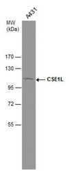
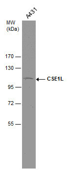
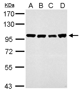
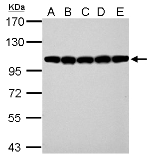
- CSE1L antibody detects CSE1L protein by western blot analysis.A. 30 ?g 293T whole cell lysate/extractB. 30 ?g A431 whole cell lysate/extractC. 30 ?g HeLa whole cell lysate/extractD. 30 ?g HepG2 whole cell lysate/extractE. 30 ?g A375 whole cell lysate/extract7.5% SDS-PAGECSE1L antibody (GTX103005) dilution: 1:10000The HRP-conjugated anti-rabbit IgG antibody (GTX213110-01) was used to detect the primary antibody.
- Submitted by
- GeneTex (provider)
- Main image

- Experimental details
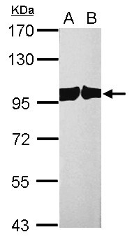
- CSE1L antibody detects CSE1L protein by western blot analysis.A. 30 ?g PC-12 whole cell lysate/extractB. 30 ?g Rat2 whole cell lysate/extract7.5% SDS-PAGECSE1L antibody (GTX103005) dilution: 1:10000The HRP-conjugated anti-rabbit IgG antibody (GTX213110-01) was used to detect the primary antibody.
- Submitted by
- GeneTex (provider)
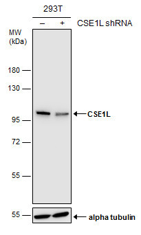
- Main image
- Experimental details
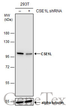
- Non-transfected (¡V) and transfected (+) 293T whole cell extracts (15 ?g) were separated by 7.5% SDS-PAGE, and the membrane was blotted with CSE1L antibody (GTX103005) diluted at 1:5000. The HRP-conjugated anti-rabbit IgG antibody (GTX213110-01) was used to detect the primary antibody.
- Submitted by
- GeneTex (provider)
- Main image
- Experimental details
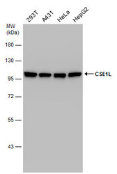
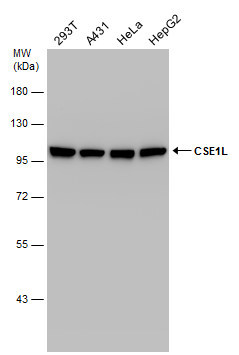
- Various whole cell extracts (30 ?g) were separated by 7.5% SDS-PAGE, and the membrane was blotted with CSE1L antibody (GTX103005) diluted at 1:10000.
Supportive validation
- Submitted by
- GeneTex (provider)
- Main image
- Experimental details
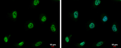
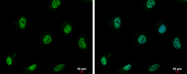
- CSE1L antibody detects CSE1L protein at nucleus by immunofluorescent analysis.Sample: HeLa cells were fixed in 4% paraformaldehyde at RT for 15 min.Green: CSE1L protein stained by CSE1L antibody (GTX103005) diluted at 1:500.Blue: Hoechst 33342 staining.Scale bar = 10 £gm.
- Submitted by
- GeneTex (provider)
- Main image

- Experimental details
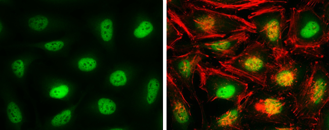
- CSE1L antibody detects CSE1L protein at nucleus by immunofluorescent analysis.Sample: HeLa cells were fixed in 4% paraformaldehyde at RT for 15 min.Green: CSE1L protein stained by CSE1L antibody (GTX103005) diluted at 1:2000.Red: phalloidin, a cytoskeleton marker, diluted at 1:100.
Supportive validation
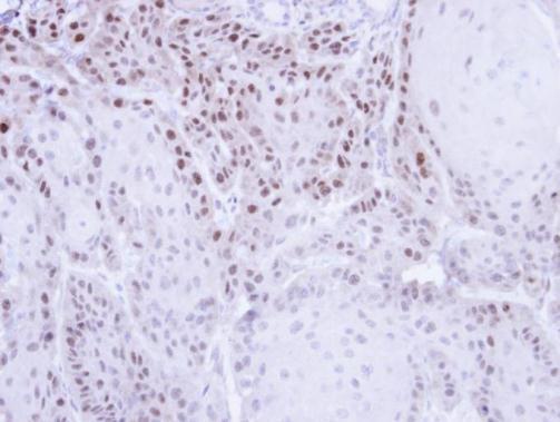
- Submitted by
- GeneTex (provider)
- Main image

- Experimental details
- Immunohistochemical analysis of paraffin-embedded Cal27 Xenograft , using CSE1L(GTX103005) antibody at 1:500 dilution.