 Explore
 Validate
 Learn
LearnPA5-21072
antibody from Invitrogen Antibodies
Targeting: SLC39A7
D6S2244E, H2-KE4, HKE4, KE4, RING5, ZIP7
 Western blot
Antibody data
- Antibody Data
- Antigen structure
- References [1]
- Comments [0]
- Validations
- Western blot [5]
- Immunohistochemistry [2]
Submit
Validation data
Reference
Comment
Report error
- Product number
- PA5-21072 - Provider product page

- Provider
- Invitrogen Antibodies
- Product name
- SLC39A7 Polyclonal Antibody
- Antibody type
- Polyclonal
- Antigen
- Synthetic peptide
- Description
- A suggested positive control is mouse brain tissue lysate.
- Concentration
- 1 mg/mL
Submitted references Hyperglycemia-Induced Changes in ZIP7 and ZnT7 Expression Cause Zn(2+) Release From the Sarco(endo)plasmic Reticulum and Mediate ER Stress in the Heart.
Tuncay E, Bitirim VC, Durak A, Carrat GRJ, Taylor KM, Rutter GA, Turan B
Diabetes 2017 May;66(5):1346-1358
Diabetes 2017 May;66(5):1346-1358
No comments: Submit comment
Supportive validation
- Submitted by
- Invitrogen Antibodies (provider)
- Main image
- Experimental details
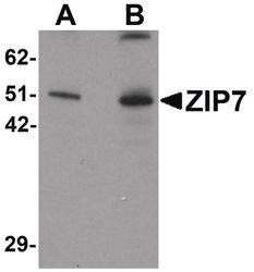
- Western blot analysis of mouse brain tissue lysate using a ZIP7 polyclonal antibody (Product # PA5-21072) at (A) 0.5 and (B) 1 µg/mL.
- Submitted by
- Invitrogen Antibodies (provider)
- Main image

- Experimental details
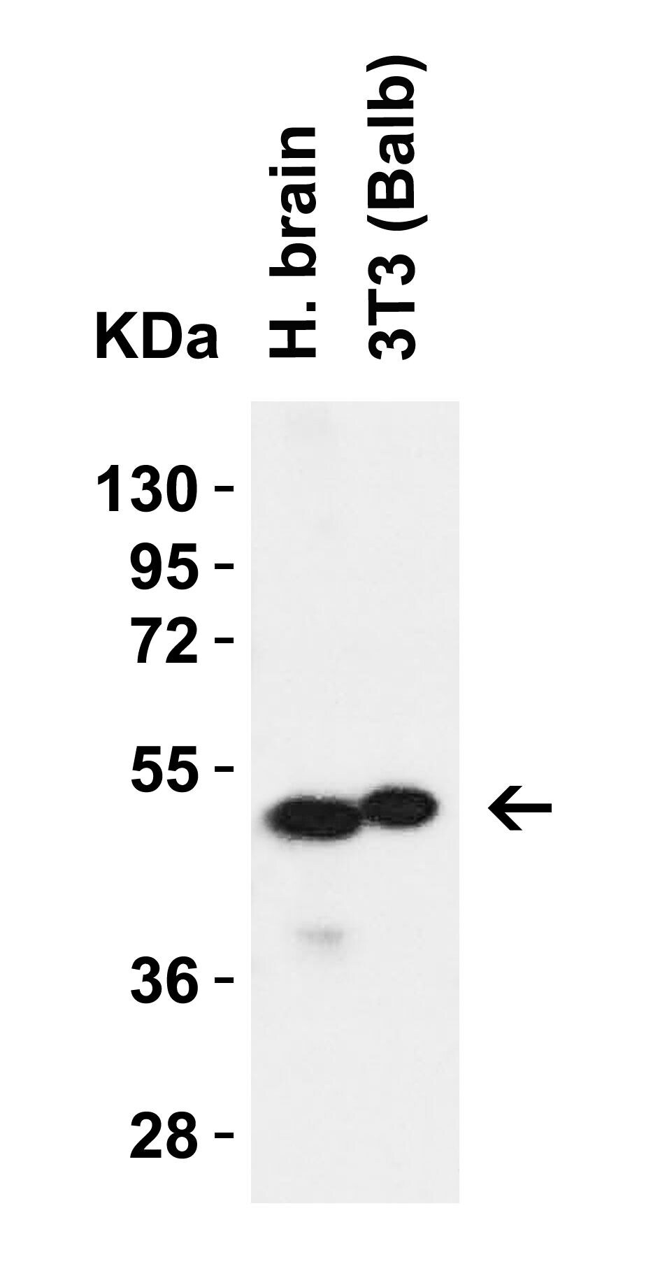
- Western Blot Validation in Human Brain Tissue and 3T3 (Balb) Cell Lysate. Loading: 15 µg of lysates per lane. Antibodies: SLC39A7 Polyclonal Antibody (Product # PA5-21072) (1 µg/mL), 1h incubation at RT in 0.05 NFDM/TBST. Secondary: Goat anti-rabbit IgG HRP conjugate at 1:10,000 dilution.
- Submitted by
- Invitrogen Antibodies (provider)
- Main image

- Experimental details
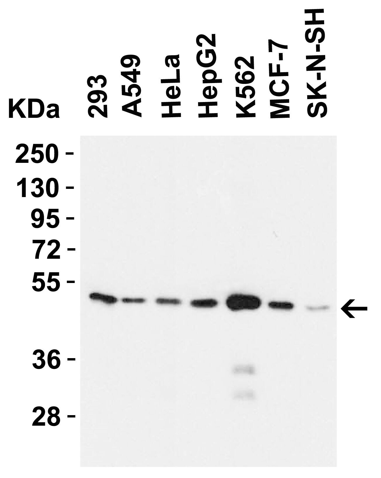
- Western Blot Validation in Human Cell Lines. Loading: 15 µg of lysates per lane. Antibodies: SLC39A7 Polyclonal Antibody (Product # PA5-21072) (1 µg/mL), 1h incubation at RT in 0.05 NFDM/TBST. Secondary: Goat anti-rabbit IgG HRP conjugate at 1:10,000 dilution.
- Submitted by
- Invitrogen Antibodies (provider)
- Main image
- Experimental details
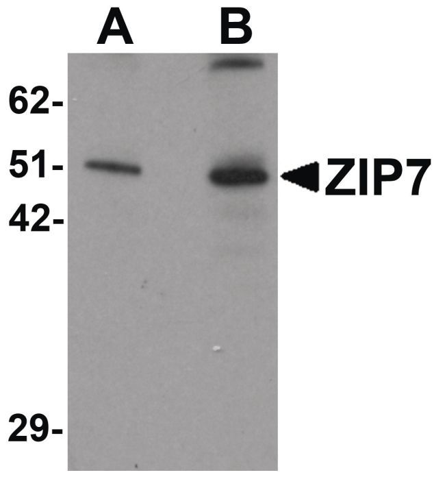
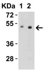
- Western Blot Validation in Mouse Brain Tissue Lysate. Loading: 15 µg of lysates per lane. Antibodies: SLC39A7 Polyclonal Antibody (Product # PA5-21072) (Lane 1: 0.5 µg/mL and Lane 2: 1 µg/mL), 1h incubation at RT in 0.05 NFDM/TBST. Secondary: Goat anti-rabbit IgG HRP conjugate at 1:10,000 dilution.

- Submitted by
- Invitrogen Antibodies (provider)
- Main image
- Experimental details
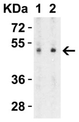
- Western Blot Validation in Rat Brain Tissue Lysate. Loading: 15 µg of lysates per lane. Antibodies: SLC39A7 Polyclonal Antibody (Product # PA5-21072) (Lane 1: 0.5 µg/mL and Lane 2: 1 µg/mL), 1h incubation at RT in 0.05 NFDM/TBST. Secondary: Goat anti-rabbit IgG HRP conjugate at 1:10,000 dilution.

Supportive validation
- Submitted by
- Invitrogen Antibodies (provider)
- Main image
- Experimental details
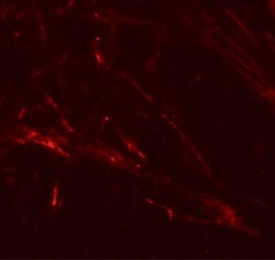
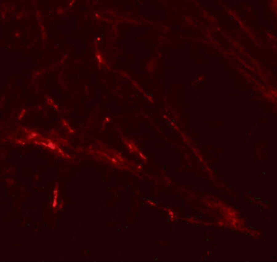
- Immunofluorescent analysis of 4% paraformaldehyde-fixed human brain tissue labeling ZIP7 with SLC39A7 Polyclonal Antibody (Product # PA5-21072) at 20 µg/mL, followed by goat anti-rabbit IgG secondary antibody at 1:500 dilution (red).
- Submitted by
- Invitrogen Antibodies (provider)
- Main image

- Experimental details
- Immunohistochemical analysis of paraffin-embedded human brain tissue using SLC39A7 Polyclonal Antibody (Product # PA5-21072) at 2.5 µg/mL. Tissue was fixed with formaldehyde and blocked with 0.1 serum for 1 h at RT; antigen retrieval was by heat mediation with a citrate buffer (pH6). Samples were incubated with primary antibody overnight at 4˚C. A goat anti-rabbit IgG H&L (HRP) at 1/250 was used as secondary. Counter stained with Hematoxylin.
