Explore
 Validate
 Learn
Learn Western blot
Antibody data
- Antibody Data
- Antigen structure
- References [0]
- Comments [0]
- Validations
- Western blot [4]
- Immunocytochemistry [2]
Submit
Validation data
Reference
Comment
Report error
- Product number
- PA5-78007 - Provider product page

- Provider
- Invitrogen Antibodies
- Product name
- HMGA1 Polyclonal Antibody
- Antibody type
- Polyclonal
- Antigen
- Recombinant full-length protein
- Description
- Positive Control: 293T, A431, HeLa, HepG2
- Concentration
- 0.87 mg/mL
No comments: Submit comment
Supportive validation
- Submitted by
- Invitrogen Antibodies (provider)
- Main image
- Experimental details
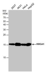
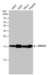
- Western blot analysis of HMGA1 in whole cell lysate using 30 µg of protein. Samples were separated with 15% SDS-PAGE and incubated with HMGA1 polyclonal antibody (Product # PA5-78007) using a dilution of 1:1000.

- Submitted by
- Invitrogen Antibodies (provider)
- Main image
- Experimental details
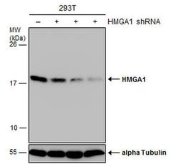
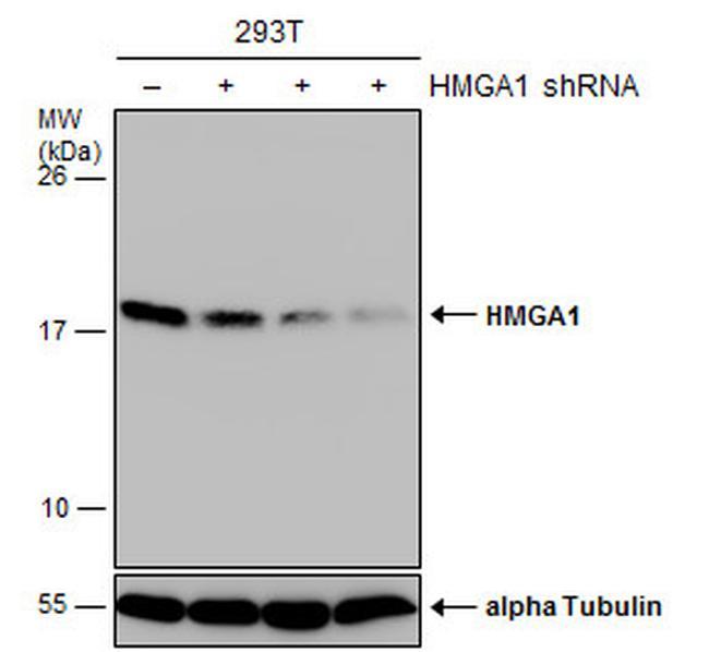
- Western blot analysis of HMGA1 in non-transfected (-) and transfected (+) 293T cells using 30 µg of protein. Samples were separated with 15% SDS-PAGE and incubated with HMGA1 polyclonal antibody (Product # PA5-78007) using a dilution of 1:3000.
- Submitted by
- Invitrogen Antibodies (provider)
- Main image
- Experimental details
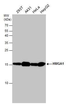
- Western Blot using HMGA1 Polyclonal Antibody (Product # PA5-78007). Various whole cell extracts (30 µg) were separated by 15% SDS-PAGE, and the membrane was blotted with HMGA1 Polyclonal Antibody (Product # PA5-78007) diluted at 1:1,000.
- Submitted by
- Invitrogen Antibodies (provider)
- Main image
- Experimental details
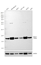
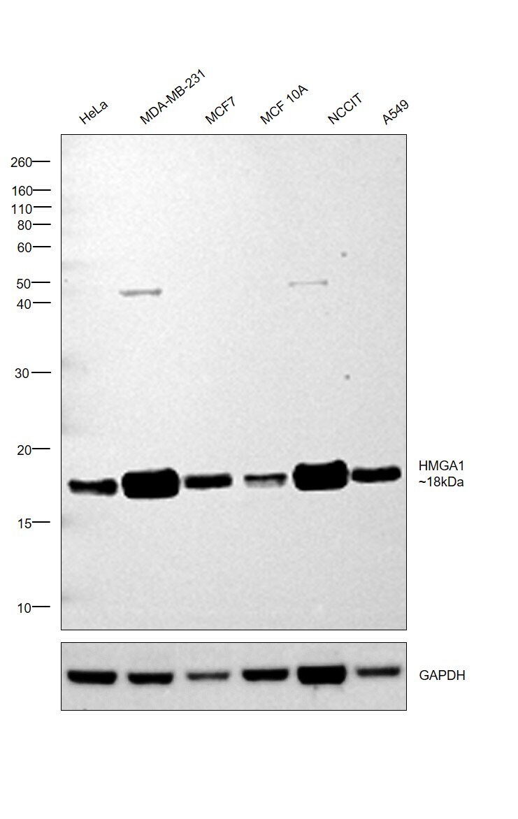
- Western blot was performed using Anti-HMGA1 Polyclonal Antibody (Product # PA5-78007) and an 18 kDa band corresponding to HMGA1 was observed in all cell lysates tested. Expression of HMGA1 correlated with the tumorigenic potential of breast cancer cell lines with higher expression being observed for MDA-MB-231 as compared to MCF7 and MCF 10A [10.1128/MCB.21.2.575-594.2001]. Modified whole cell extracts (1% SDS) (30 µg lysate) of HeLa (Lane 1), MDA-MB-231 (Lane 2), MCF7 (Lane 3), MCF 10A (Lane 4), NCCIT (Lane 5) and A549 (Lane 6) were electrophoresed using Novex® NuPAGE® 4-12 % Bis-Tris gel (Product # NP0322BOX). Resolved proteins were then transferred onto a nitrocellulose membrane (Product # IB23001) by iBlot® 2 Dry Blotting System (Product # IB21001). The blot was probed with the primary antibody (1:1000 dilution) and detected by chemiluminescence with Goat anti-Rabbit IgG (H+L), Superclonal™ Recombinant Secondary Antibody, HRP (Product # A27036, 1:4000 dilution) using the iBright FL 1000 (Product # A32752). Chemiluminescent detection was performed using Novex® ECL Chemiluminescent Substrate Reagent Kit (Product # WP20005).
Supportive validation
- Submitted by
- Invitrogen Antibodies (provider)
- Main image
- Experimental details
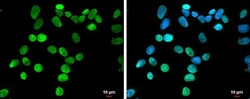
- HMGA1 Polyclonal Antibody detects HMGA1 protein at nucleus by immunofluorescent analysis. Sample: MCF7 cells were fixed in 4% paraformaldehyde at RT for 15 min. Green: HMGA1 protein stained by HMGA1 Polyclonal Antibody (Product # PA5-78007) diluted at 1:500. Blue: Hoechst 33342 staining. Scale bar = 10 µm.
- Submitted by
- Invitrogen Antibodies (provider)
- Main image
- Experimental details
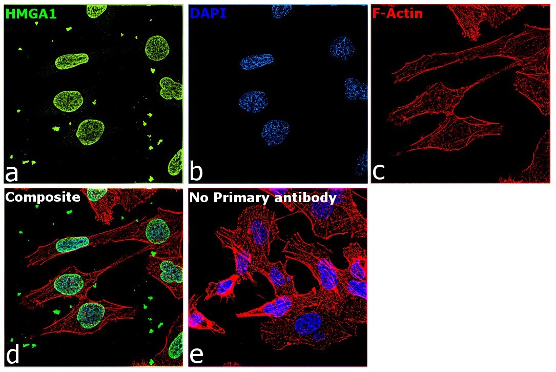
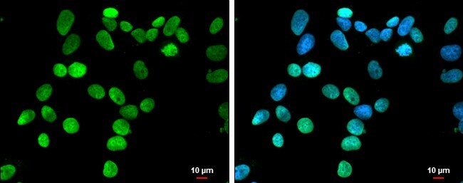
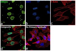
- Immunofluorescence analysis of HMGA1 was performed using 70% confluent log phase MDA-MB-231 cells. The cells were fixed with 4% paraformaldehyde for 10 minutes, permeabilized with 0.1% Triton™ X-100 for 15 minutes, and blocked with 2% BSA for 1 hour at room temperature. The cells were labeled with HMGA1 Polyclonal Antibody (Product # PA5-78007) at 1:100 dilution in 0.1% BSA, incubated at 4 degree Celsius overnight and then labeled with Donkey anti-Rabbit IgG (H+L) Highly Cross-Adsorbed Secondary Antibody, Alexa Fluor Plus 488 (Product # A32790) at a dilution of 1:2000 for 45 minutes at room temperature (Panel a: green). Nuclei (Panel b: blue) were stained with SlowFade® Gold Antifade Mountant with DAPI (Product # S36938). F-actin (Panel c: red) was stained with Rhodamine Phalloidin (Product # R415, 1:300). Panel d represents the merged image showing localization to nucleus. Panel f represents control cells with no primary antibody to assess background. The images were captured at 60X magnification.