 Explore
 Validate
 Learn
Learn Western blot
Antibody data
- Antibody Data
- Antigen structure
- References [0]
- Comments [0]
- Validations
- Western blot [1]
- Immunocytochemistry [1]
- Flow cytometry [1]
Submit
Validation data
Reference
Comment
Report error
- Product number
- MAB4734 - Provider product page

- Provider
- R&D Systems
- Product name
- Human TSPAN8 Antibody
- Antibody type
- Monoclonal
- Description
- Protein A or G purified from hybridoma culture supernatant. Detects human TSPAN8 in Western blots.
- Reactivity
- Human
- Host
- Rat
- Conjugate
- Unconjugated
- Antigen sequence
AAH05246- Isotype
- IgG
- Antibody clone number
- 458811
- Vial size
- 100 ug
- Concentration
- LYOPH
- Storage
- Use a manual defrost freezer and avoid repeated freeze-thaw cycles. 12 months from date of receipt, -20 to -70 °C as supplied. 1 month, 2 to 8 °C under sterile conditions after reconstitution. 6 months, -20 to -70 °C under sterile conditions after reconstitution.
No comments: Submit comment
Supportive validation
- Submitted by
- R&D Systems (provider)
- Main image
- Experimental details
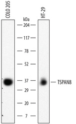
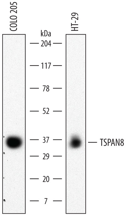
- Detection of Human TSPAN8 by Western Blot. Western blot shows lysates of COLO 205 human colorectal adenocarcinoma cell line and HT-29 human colon adenocarcinoma cell line. PVDF membrane was probed with 2 µg/mL of Rat Anti-Human TSPAN8 Monoclonal Antibody (Catalog # MAB4734) followed by HRP-conjugated Anti-Rat IgG Secondary Antibody (Catalog # HAF005). A specific band was detected for TSPAN8 at approximately 35 kDa (as indicated). This experiment was conducted under non-reducing conditions and using Immunoblot Buffer Group 1.
Supportive validation
- Submitted by
- R&D Systems (provider)
- Main image

- Experimental details
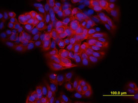
- TSPAN8 in HT-29 Human Cell Line. TSPAN8 was detected in immersion fixed HT-29 human colon adenocarcinoma cell line using Rat Anti-Human TSPAN8 Monoclonal Antibody (Catalog # MAB4734) at 10 µg/mL for 3 hours at room temperature. Cells were stained using the NorthernLights™ 557-conjugated Anti-Rat IgG Secondary Antibody (red; Catalog # NL013) and counterstained with DAPI (blue). View our protocol for Fluorescent ICC Staining of Cells on Coverslips.
Supportive validation
- Submitted by
- R&D Systems (provider)
- Main image
- Experimental details
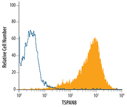
- Detection of TSPAN8 in HT-29 Human Cell Line by Flow Cytometry. HT-29 human colon adenocarcinoma cell line was stained with Rat Anti-Human TSPAN8 Monoclonal Antibody (Catalog # MAB4734, filled histogram) or isotype control antibody (Catalog # MAB0061, open histogram), followed by Allophycocyanin-conjugated Anti-Rat IgG F(ab')2 Secondary Antibody (Catalog # F0113).
