Explore
 Validate
 Learn
LearnGTX23446
antibody from GeneTex
Targeting: NFAT5
KIAA0827, NF-AT5, NFATL1, NFATZ, OREBP, TONEBP
 Western blot Immunocytochemistry Immunoprecipitation
 Immunohistochemistry Gel shift Chromatin Immunoprecipitation
Antibody data
- Antibody Data
- Antigen structure
- References [0]
- Comments [0]
- Validations
- Western blot [2]
- Immunocytochemistry [1]
- Immunoprecipitation [1]
Submit
Validation data
Reference
Comment
Report error
- Product number
- GTX23446 - Provider product page

- Provider
- GeneTex
- Proper citation
- GeneTex Cat#GTX23446, RRID:AB_385052
- Product name
- NFAT5 antibody
- Antibody type
- Polyclonal
- Reactivity
- Human, Mouse, Rat, Rabbit, Simian
- Host
- Rabbit
No comments: Submit comment
Supportive validation
- Submitted by
- GeneTex (provider)
- Main image
- Experimental details
- Western blot analysis of NFAT5 in 25ug of various whole cell lysates. Proteins were transferred to a PVDF membrane and blocked with 5% Milk/TBST for at least 1 hour. Membranes were probed with NFAT5 antibody at a dilution of 1:1000 overnight at 4¢XC on a rocking platform. Membranes were washed in TBS-0.1%Tween 20 and probed with a HRP-conjugated secondary antibody. Membranes were washed and chemiluminescent detection performed.

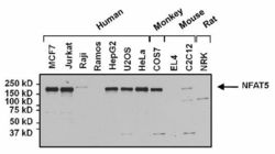
- Submitted by
- GeneTex (provider)
- Main image
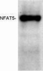
- Experimental details
- Western blot of human NFAT5 from transfected BHK cell lysate with GTX23446.
- Validation comment
- WB

Supportive validation
- Submitted by
- GeneTex (provider)
- Main image
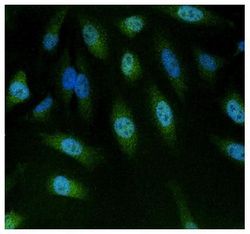
- Experimental details
- Immunofluorescent analysis of NFAT5 in HeLa cells. Formalin-fixed cells were permeabilized with 0.1% Triton X-100 in TBS for 10 minutes at room temperature. Cells were then blocked with 1% BSA for 15 minutes at room temperature. Cells were probed with NFAT5 antibody at a dilution of 1:100 for at least 1 hour at room temperature. Cells were washed with PBS and incubated with a proper secondary antibody. Nuclei (blue) were stained with Hoechst 33342 dye. Images were taken at 20X magnification.

Supportive validation
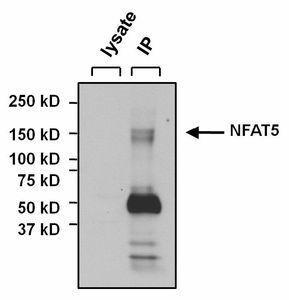
- Submitted by
- GeneTex (provider)
- Main image

- Experimental details
- Immunoprecipitation of NFAT5frpm U2OS cells. The antigen-antibody complex was formed by incubating 500£gg whole cell lysate with 3£gg of NFAT5 antibody overnight on a rocking platform at 4¢XC. The immune-complex was captured on 50£gl Protein A/G Agarose. Captured immune-complexes were washed and eluted. Samples were resolved on a 4-20% Tris-HCl polyacrylamide gel. Proteins were transferred to PVDF membrane and blocked with 5% Milk/TBS-0.1%Tween for at least 1 hour. Membranes were washed in TBS-0.1%Tween 20 and probed with a proper secondary antibody. Membranes were washed and chemiluminescent detection performed.