Explore
 Validate
 Learn
LearnSP5110P
antibody from Acris Antibodies GmbH
Targeting: NFAT5
KIAA0827, NF-AT5, NFATL1, NFATZ, OREBP, TONEBP
 Western blot
 Immunoelectron microscopy
Antibody data
- Antibody Data
- Antigen structure
- References [0]
- Comments [0]
- Validations
- Western blot [2]
- Immunocytochemistry [1]
- Immunoprecipitation [1]
Submit
Validation data
Reference
Comment
Report error
- Product number
- SP5110P - Provider product page

- Provider
- Acris Antibodies GmbH
- Proper citation
- Acris Antibodies GmbH Cat#SP5110P, RRID:AB_1005663
- Product name
- anti NFAT5
- Antibody type
- Polyclonal
- Antigen
- Synthetic peptide corresponding to residues 1439-1455 of Human NFAT5.
- Reactivity
- Human, Mouse, Rat, Porcine
- Host
- Rabbit
- Isotype
- IgG
- Vial size
- 50 µg
- Concentration
- 1.0 mg/ml
No comments: Submit comment
Supportive validation
- Submitted by
- Acris Antibodies GmbH (provider)
- Main image

- Experimental details
- Figure 1. Western blot of Human NFAT5 from transfected BHK cell lysate with SP5110P.

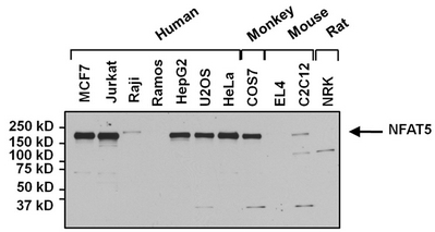
- Submitted by
- Acris Antibodies GmbH (provider)
- Main image

- Experimental details
- Figure 4. Western blot analysis of NFAT5 was performed by loading 25 µg of various whole cell lysates onto a 4-20% Tris-HCl polyacrylamide gel. Proteins were transferred to a PVDF membrane and blocked with 5% Milk/TBST for at least 1 hour. Membranes were probed with a Rabbit Polyclonal antibody recognizing NFAT5 (SP5110P) at a dilution of 1/1000 overnight at 4°C on a rocking platform. Membranes were washed in TBS-0.1%Tween 20 and probed with a Goat anti-Rabbit-HRP secondary antibody at a dilution of 1/20,000 for at least one hour. Membranes were washed and chemiluminescent detection performed.
Supportive validation
- Submitted by
- Acris Antibodies GmbH (provider)
- Main image
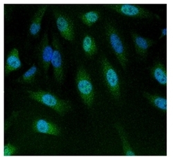
- Experimental details
- Figure 2. Immunofluorescent analysis of NFAT5 using SP5110P NFAT5 antibody (shown in Green) in HeLa cells. Formalin fixed cells were permeabilized with 0.1% Triton X-100 in TBS for 10 minutes at RT. Cells were then blocked with 1% Blocker BSA for 15 minutes at RT. Cells were probed with a Rabbit Polyclonal Antibody recognizing NFAT5 (SP5110P), at a dilution of 1/100 for at least 1 hour at RT. Cells were washed with PBS and incubated with DyLight 488 Goat-anti-Rabbit secondary antibody at a dilution of 1/400 for 30 minutes at RT. Nuclei (blue) were stained with Hoechst 33342 dye. Images were taken on a ArrayScan at 20X magnification.

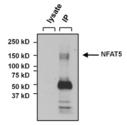
Supportive validation
- Submitted by
- Acris Antibodies GmbH (provider)
- Main image

- Experimental details
- Figure 3. Immunoprecipitation of NFAT5 was performed on U2OS cells. The antigen:antibody complex was formed by incubating 500µg whole cell lysate with 3µg of Rabbit Polyclonal antibody recognizing NFAT5 overnight on a rocking platform at 4°C. The immune-complex was captured on 50µl Protein A/G Plus Agarose. Captured immune-complexes were washed and proteins eluted with 5X Reducing Sample Loading Dye. Samples were resolved on a 4-20% Tris-HCl polyacrylamide gel. Proteins were transferred to PVDF membrane and blocked with 5% Milk/TBS-0.1%Tween for at least 1 hour. Membranes were washed in TBS-0.1%Tween 20 and probed with a Goat anti-Rabbit-HRP secondary antibody at a dilution of 1/20,000 for at least one hour. Membranes were washed and chemiluminescent detection performed using Super Signal West Dura.