 Explore
 Validate
 Learn
LearnPA5-27593
antibody from Invitrogen Antibodies
Targeting: EIF3K
ARG134, EIF3S12, HSPC029, M9, PLAC-24, PRO1474, PTD001
 Western blot
Antibody data
- Antibody Data
- Antigen structure
- References [1]
- Comments [0]
- Validations
- Western blot [1]
- Immunocytochemistry [1]
- Immunohistochemistry [2]
- Other assay [2]
Submit
Validation data
Reference
Comment
Report error
- Product number
- PA5-27593 - Provider product page

- Provider
- Invitrogen Antibodies
- Product name
- eIF3k Polyclonal Antibody
- Antibody type
- Polyclonal
- Antigen
- Recombinant protein fragment
- Description
- Recommended positive controls: 293T.
- Concentration
- 10 mg/mL
Submitted references New Proteins Contributing to Immune Cell Infiltration and Pannus Formation of Synovial Membrane from Arthritis Diseases.
de Seny D, Baiwir D, Bianchi E, Cobraiville G, Deroyer C, Poulet C, Malaise O, Paulissen G, Kaiser MJ, Hauzeur JP, Mazzucchelli G, Delvenne P, Malaise M
International journal of molecular sciences 2021 Dec 31;23(1)
International journal of molecular sciences 2021 Dec 31;23(1)
No comments: Submit comment
Supportive validation
- Submitted by
- Invitrogen Antibodies (provider)
- Main image

- Experimental details
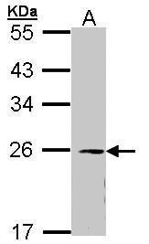
- Western Blot using eIF3k Polyclonal Antibody (Product # PA5-27593). Sample (30 µg of whole cell lysate). Lane A: 293T. 12% SDS PAGE. EIF3k Polyclonal Antibody (Product # PA5-27593) diluted at 1:1,000.
Supportive validation
- Submitted by
- Invitrogen Antibodies (provider)
- Main image
- Experimental details
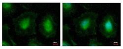
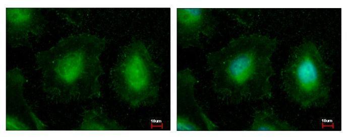
- eIF3k Polyclonal Antibody detects EIF3K protein at cytoplasm and nucleus by immunofluorescent analysis. Sample: HeLa cells were fixed in 4% paraformaldehyde at RT for 15 min. Green: EIF3K protein stained by eIF3k Polyclonal Antibody (Product # PA5-27593) diluted at 1:500. Blue: Hoechst 33343 staining.
Supportive validation
- Submitted by
- Invitrogen Antibodies (provider)
- Main image
- Experimental details
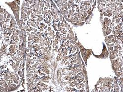
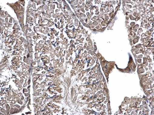
- eIF3k Polyclonal Antibody detects EIF3K protein at cytosol on mouse testis by immunohistochemical analysis. Sample: Paraffin-embedded mouse testis. EIF3k Polyclonal Antibody (Product # PA5-27593) dilution: 1:500. Antigen Retrieval: EDTA based buffer, pH 8.0, 15 min.
- Submitted by
- Invitrogen Antibodies (provider)
- Main image
- Experimental details
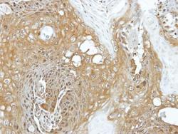
- Immunohistochemical analysis of paraffin-embedded Cal27 xenograft, using EIF3K (Product # PA5-27593) antibody at 1:100 dilution. Antigen Retrieval: EDTA based buffer, pH 8.0, 15 min.

Supportive validation
- Submitted by
- Invitrogen Antibodies (provider)
- Main image
- Experimental details
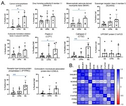
- Figure 2 Immunohistochemistry (IHC) quantification of the 10 highlighted proteins in synovial membrane from OA, CPPA and RA patients. ( A ) Representation of protein quantification (optical density values) obtained by QuPath after IHC. One-way ANOVA test (post hoc of Tukey) or Kruskal-Wallis test (post hoc test of Dunn's) was applied depending on normal distribution: * P < 0.05 and ** P < 0.01 ( B ) Correlation coefficients between the 10 highlighted biomarkers calculated according to the non-parametric Spearman test. OA, osteoarthritis; CPPA, chronic pyrophosphate arthropathy; RA, rheumatoid arthritis.

- Submitted by
- Invitrogen Antibodies (provider)
- Main image
- Experimental details
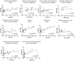
- Figure 3 Correlation between mass spectrometry (MS) and immunohistochemistry (IHC) quantification of the 10 highlighted proteins in synovial membrane from OA, CPPA and RA patients. Protein expression levels (Log2 (LFQ)) obtained by mass spectrometry were correlated to the percentage of positive cells obtained by IHC using the non-parametric Spearman test: * P < 0.05, ** P < 0.01 and *** P < 0.001.
