Explore
 Validate
 Learn
Learn Western blot
 Immunohistochemistry
Antibody data
- Antibody Data
- Antigen structure
- References [0]
- Comments [0]
- Validations
- Western blot [2]
- Immunocytochemistry [1]
- Immunohistochemistry [6]
Submit
Validation data
Reference
Comment
Report error
- Product number
- HPA047834 - Provider product page

- Provider
- Atlas Antibodies
- Proper citation
- Atlas Antibodies Cat#HPA047834, RRID:AB_2680172
- Product name
- Anti-BHLHA15
- Antibody type
- Polyclonal
- Reactivity
- Human
- Host
- Rabbit
- Conjugate
- Unconjugated
- Antigen sequence
AKNYIKSLTATILTMSSSRLPGLEGPGPKLYQHYQ
QQQQVAGGALGATEAQPQGHLQRYSTQIHSFREG- Isotype
- IgG
- Vial size
- 100 µl
- Storage
- Store at +4°C for short term storage. Long time storage is recommended at -20°C.
No comments: Submit comment
Supportive validation
- Submitted by
- Atlas Antibodies (provider)
- Enhanced method
- Recombinant expression validation
- Main image
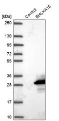
- Experimental details
- Western blot analysis in control (vector only transfected HEK293T lysate) and BHLHA15 over-expression lysate (Co-expressed with a C-terminal myc-DDK tag (~3.1 kDa) in mammalian HEK293T cells, LY406171).
- Submitted by
- Atlas Antibodies (provider)
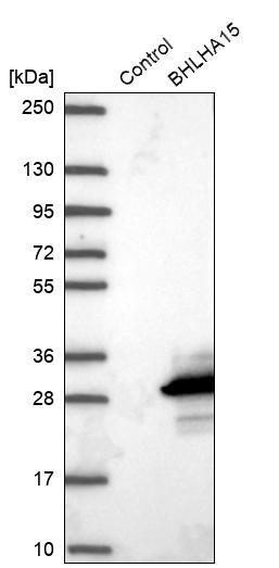
- Main image

- Experimental details
- Western blot analysis in human cell line RPMI-8226.

Supportive validation
- Submitted by
- Atlas Antibodies (provider)
- Main image
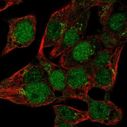
- Experimental details
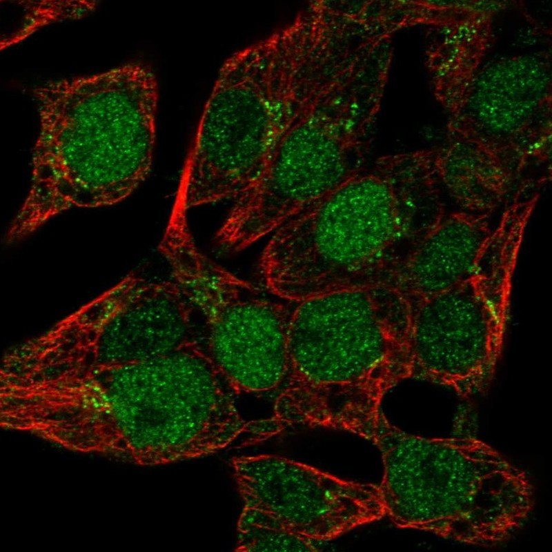
- Immunofluorescent staining of human cell line Hep G2 shows localization to nucleoplasm & the Golgi apparatus.
- Sample type
- HUMAN
Enhanced validation
Supportive validation
- Submitted by
- Atlas Antibodies (provider)
- Enhanced method
- Orthogonal validation
- Main image
- Experimental details
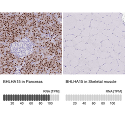
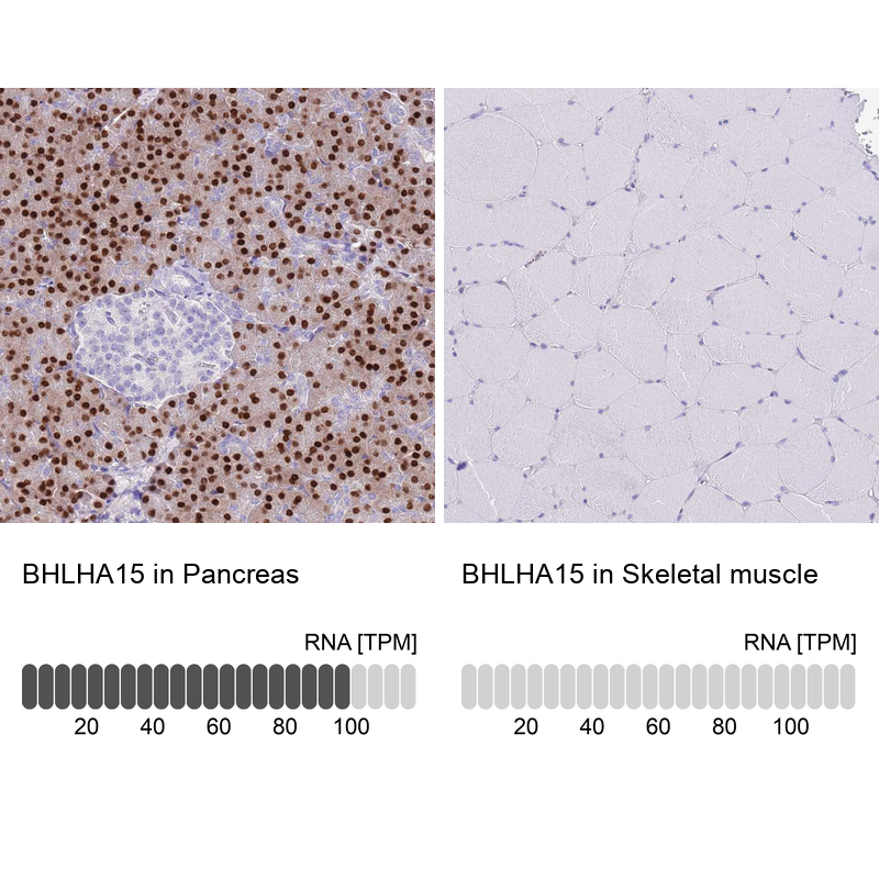
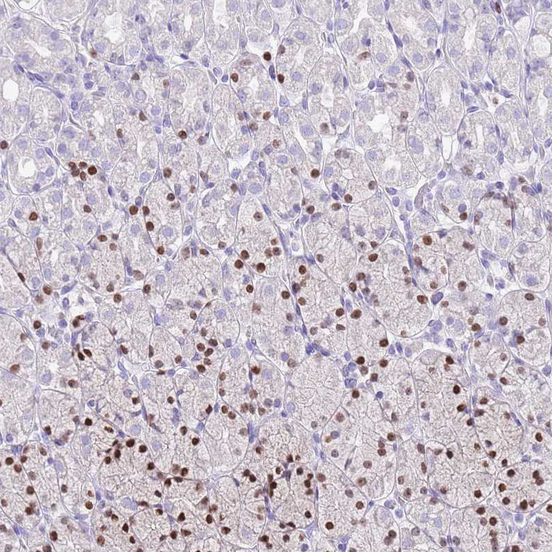
- Immunohistochemistry analysis in human pancreas and skeletal muscle tissues using HPA047834 antibody. Corresponding BHLHA15 RNA-seq data are presented for the same tissues.
- Sample type
- HUMAN
Supportive validation
- Submitted by
- Atlas Antibodies (provider)
- Main image
- Experimental details
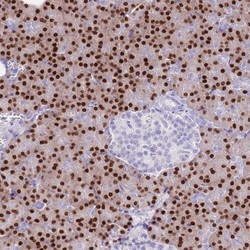
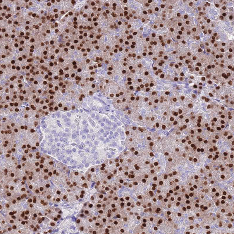
- Immunohistochemical staining of human pancreas shows strong nuclear positivity in exocrine glandular cells.

- Submitted by
- Atlas Antibodies (provider)
- Main image

- Experimental details
- Immunohistochemical staining of human pancreas shows strong nuclear positivity in exocrine glandular cells.
- Sample type
- HUMAN
- Submitted by
- Atlas Antibodies (provider)
- Main image
- Experimental details
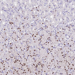
- Immunohistochemical staining of human stomach shows moderate to strong nuclear positivity in a subset of glandular cells.
- Sample type
- HUMAN
- Submitted by
- Atlas Antibodies (provider)
- Main image
- Experimental details
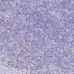
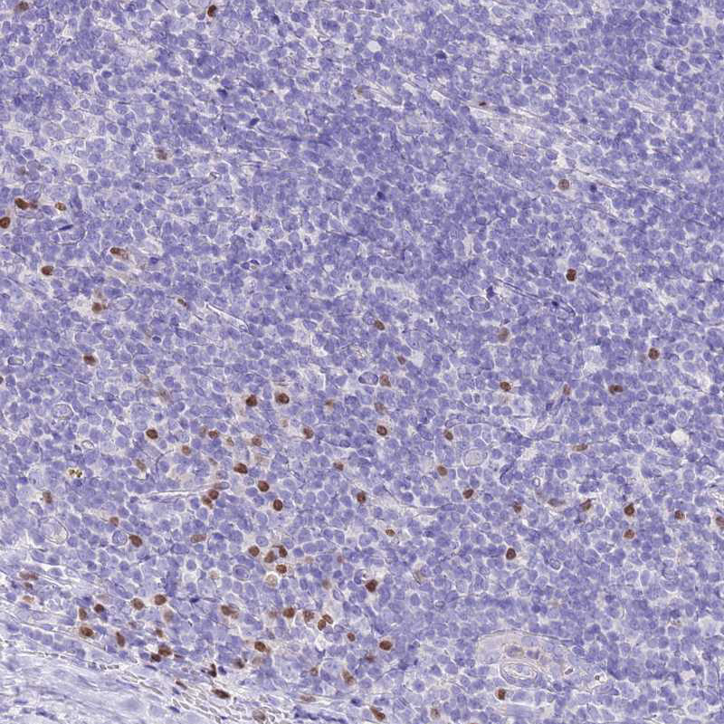
- Immunohistochemical staining of human tonsil shows moderate to strong nuclear positivity in non-germinal center cells.
- Sample type
- HUMAN
- Submitted by
- Atlas Antibodies (provider)
- Main image
- Experimental details
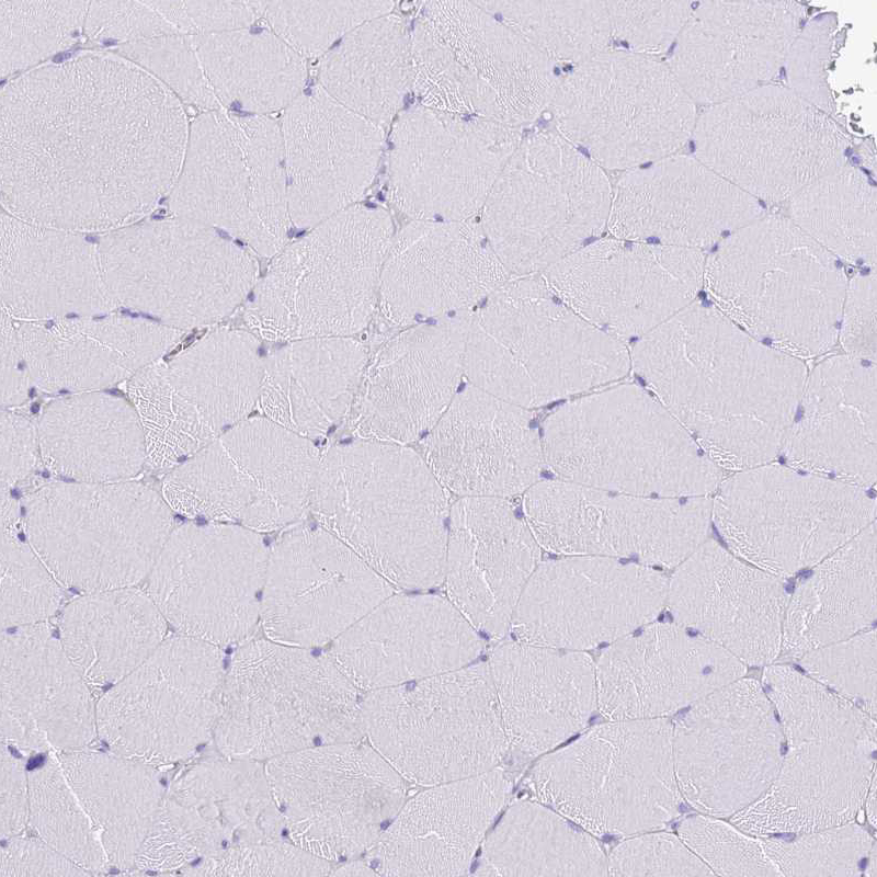
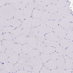
- Immunohistochemical staining of human skeletal muscle shows no positivity in striated muscle fibers as expected.
- Sample type
- HUMAN