 Explore
 Validate
 Learn
Learn Western blot
Antibody data
- Antibody Data
- Antigen structure
- References [3]
- Comments [0]
- Validations
- Western blot [2]
- Other assay [10]
Submit
Validation data
Reference
Comment
Report error
- Product number
- PA1-1961 - Provider product page

- Provider
- Invitrogen Antibodies
- Product name
- PSME4 Polyclonal Antibody
- Antibody type
- Polyclonal
- Antigen
- Synthetic peptide
- Description
- PA1-1961 detects PA200 from bovine, human, mouse and rat samples.
- Concentration
- Conc. Not Determined
Submitted references The proteasome activator PA200 regulates expression of genes involved in cell survival upon selective mitochondrial inhibition in neuroblastoma cells.
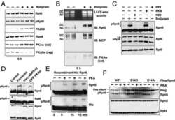
cAMP-induced phosphorylation of 26S proteasomes on Rpn6/PSMD11 enhances their activity and the degradation of misfolded proteins.
PA200, a nuclear proteasome activator involved in DNA repair.
Douida A, Batista F, Robaszkiewicz A, Boto P, Aladdin A, Szenykiv M, Czinege R, Virág L, Tar K
Journal of cellular and molecular medicine 2020 Jun;24(12):6716-6730
Journal of cellular and molecular medicine 2020 Jun;24(12):6716-6730
cAMP-induced phosphorylation of 26S proteasomes on Rpn6/PSMD11 enhances their activity and the degradation of misfolded proteins.
Lokireddy S, Kukushkin NV, Goldberg AL
Proceedings of the National Academy of Sciences of the United States of America 2015 Dec 29;112(52):E7176-85
Proceedings of the National Academy of Sciences of the United States of America 2015 Dec 29;112(52):E7176-85
PA200, a nuclear proteasome activator involved in DNA repair.
Ustrell V, Hoffman L, Pratt G, Rechsteiner M
The EMBO journal 2002 Jul 1;21(13):3516-25
The EMBO journal 2002 Jul 1;21(13):3516-25
No comments: Submit comment
Supportive validation
- Submitted by
- Invitrogen Antibodies (provider)
- Main image

- Experimental details
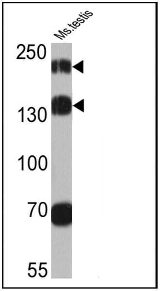
- Western blot analysis of Proteosome Activator, 200 kDa was performed by loading 25 µg of mouse testis cell lysates onto an SDS polyacrylamide gel. Proteins were transferred to a PVDF membrane and blocked at 4ºC overnight. The membrane was probed with a Proteosome Activator, 200 kDa polyclonal antibody (Product # PA1-1961) at a dilution of 1:2000 overnight at 4°C, washed in TBST, and probed with an HRP-conjugated secondary antibody for 1 hr at room temperature in the dark. Chemiluminescent detection was performed using Pierce ECL Plus Western Blotting Substrate (Product # 32132). Results show a band at ~160 and 200 kDa.
- Submitted by
- Invitrogen Antibodies (provider)
- Main image
- Experimental details
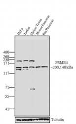
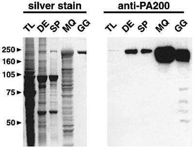
- Western blot analysis was performed on whole cell extracts (30 µg lysate) of HeLa (Lane 1), Jurkat (Lane 2), and tissue extracts (30 µg lysate) of Mouse testis (Lane 3), Mouse pancreas (Lane 4), and Rat pancreas (Lane 5). The blot was probed with Anti-PSME4 Rabbit Polyclonal Antibody (Product # PA1-1961, 1:250 dilution) and detected by chemiluminescence using Goat anti-Rabbit IgG (H+L) Superclonal™ Secondary Antibody, HRP conjugate (Product # A27036, 0.25 µg/mL, 1:4000 dilution). A 200 kDa band corresponding to PSME4 was observed in HeLa, Jurkat, and mouse testis. In addition, a 140 kDa band corresponding to an isoform of PSME4 was observed across the cell lines and tissues tested. Known quantity of protein samples were electrophoresed using Novex® NuPAGE® 4-12 % Bis-Tris gel (Product # NP0322BOX), XCell SureLock™ Electrophoresis System (Product # EI0002) and HiMark™ Pre-stained Protein Standard (Product # LC5699). Resolved proteins were then transferred onto a nitrocellulose membrane using the overnight wet transfer (tank transfer) technique. The membrane was probed with the relevant primary and secondary Antibody following blocking with 5% skimmed milk. Chemiluminescent detection was performed using Pierce™ ECL Western Blotting Substrate (Product # 32106).

Supportive validation
- Submitted by
- Invitrogen Antibodies (provider)
- Main image

- Experimental details
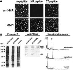
- NULL
- Submitted by
- Invitrogen Antibodies (provider)
- Main image
- Experimental details
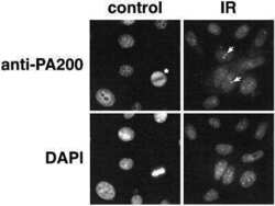
- NULL

- Submitted by
- Invitrogen Antibodies (provider)
- Main image

- Experimental details
- NULL
- Submitted by
- Invitrogen Antibodies (provider)
- Main image
- Experimental details
- NULL

- Submitted by
- Invitrogen Antibodies (provider)
- Main image
- Experimental details
- NULL

- Submitted by
- Invitrogen Antibodies (provider)
- Main image
- Experimental details
- NULL
- Submitted by
- Invitrogen Antibodies (provider)
- Main image
- Experimental details
- NULL

- Submitted by
- Invitrogen Antibodies (provider)
- Main image
- Experimental details
- NULL
- Submitted by
- Invitrogen Antibodies (provider)
- Main image
- Experimental details
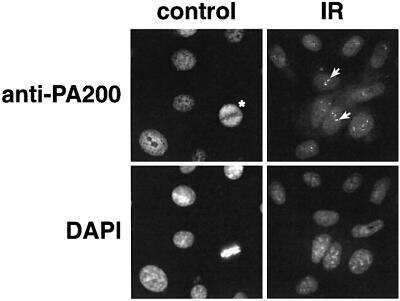
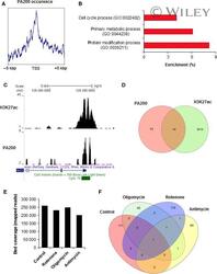
- 4 FIGURE PA200 occurrence in the genome of SH-SY5Y responds to cell treatment with mitochondria-impairing agents. (A) PA200 distribution around gene transcription start sites (TSS) was monitored by analysing mapped ChIP-seq reads with deepTools. (B) and (C) Peak calling in MACS revealed PA200 presence at gene promoters and their biological function, analysed with Amigo2 (B), disclosed crucial cellular processes. (C) Some PA200-positive gene promoters were simultaneously characterized by histone acetylation (H3K27ac) that marks actively transcribed genes. (D) Venn diagrams display PA200-enriched active (+H3K27ac) and inactive (-H3K27ac) gene promoters. (E) PA200 association with chromatin after cell treatment with 10 mumol/L rotenone, 3 mumol/L oligomycin and 100 nmol/L antimycin A was monitored and quantified by bedCoverage. (F) Alterations in the occupancy of gene promoters by PA200 in untreated cells and cells challenged with mitochondria-impairing agents were compared by Venn diagrams
- Submitted by
- Invitrogen Antibodies (provider)
- Main image
- Experimental details
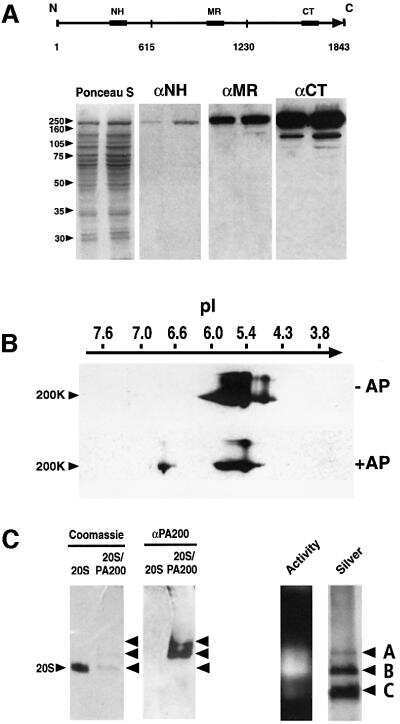
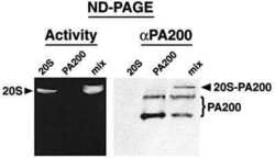
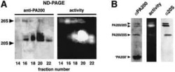
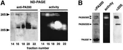
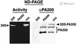
- Proteasome activation by purified bovine PA200. Purified PA200 or 20S proteasomes were analyzed separately or mixed prior to electrophoresis on a native gel. Peptide overlay (left) demonstrated that PA200 activated the proteasome and western blotting (right) confirmed that some of the added PA200 bound the 20S proteasome (denoted 20S-PA200) in the right panel.
