 Explore
 Validate
 Learn
Learn Western blot
Antibody data
- Antibody Data
- Antigen structure
- References [4]
- Comments [0]
- Validations
- Western blot [4]
- Immunocytochemistry [1]
Submit
Validation data
Reference
Comment
Report error
- Product number
- GTX110473 - Provider product page

- Provider
- GeneTex
- Proper citation
- GeneTex Cat#GTX110473, RRID:AB_10720524
- Product name
- TDG antibody
- Antibody type
- Polyclonal
- Reactivity
- Human
- Host
- Rabbit
Submitted references Characterizing Requirements for Small Ubiquitin-like Modifier (SUMO) Modification and Binding on Base Excision Repair Activity of Thymine-DNA Glycosylase in Vivo.
Minimal role of base excision repair in TET-induced global DNA demethylation in HEK293T cells.
SUMO-modification and elimination of the active DNA demethylation enzyme TDG in cultured human cells.
Thymine DNA glycosylase is a positive regulator of Wnt signaling in colorectal cancer.
McLaughlin D, Coey CT, Yang WC, Drohat AC, Matunis MJ
The Journal of biological chemistry 2016 Apr 22;291(17):9014-24
The Journal of biological chemistry 2016 Apr 22;291(17):9014-24
Minimal role of base excision repair in TET-induced global DNA demethylation in HEK293T cells.
Jin C, Qin T, Barton MC, Jelinek J, Issa JP
Epigenetics 2015;10(11):1006-13
Epigenetics 2015;10(11):1006-13
SUMO-modification and elimination of the active DNA demethylation enzyme TDG in cultured human cells.
Moriyama T, Fujimitsu Y, Yoshikai Y, Sasano T, Yamada K, Murakami M, Urano T, Sugasawa K, Saitoh H
Biochemical and biophysical research communications 2014 May 9;447(3):419-24
Biochemical and biophysical research communications 2014 May 9;447(3):419-24
Thymine DNA glycosylase is a positive regulator of Wnt signaling in colorectal cancer.
Xu X, Yu T, Shi J, Chen X, Zhang W, Lin T, Liu Z, Wang Y, Zeng Z, Wang C, Li M, Liu C
The Journal of biological chemistry 2014 Mar 28;289(13):8881-90
The Journal of biological chemistry 2014 Mar 28;289(13):8881-90
No comments: Submit comment
Supportive validation
- Submitted by
- GeneTex (provider)
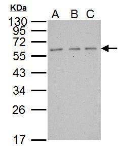
- Main image

- Experimental details
- Sample (30 ?g of whole cell lysate) A: 293T B: A431 C: HeLa 12% SDS PAGE GTX110473 diluted at 1:5000 The HRP-conjugated anti-rabbit IgG antibody (GTX213110-01) was used to detect the primary antibody.
- Submitted by
- GeneTex (provider)
- Main image
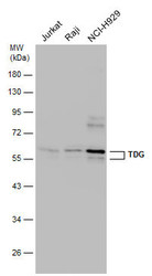
- Experimental details
- Various whole cell extracts (30 ?g) were separated by 10% SDS-PAGE, and the membrane was blotted with TDG antibody (GTX110473) diluted at 1:500. The HRP-conjugated anti-rabbit IgG antibody (GTX213110-01) was used to detect the primary antibody.
- Submitted by
- GeneTex (provider)
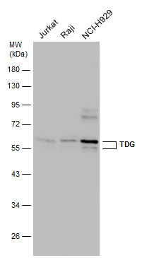
- Main image
- Experimental details
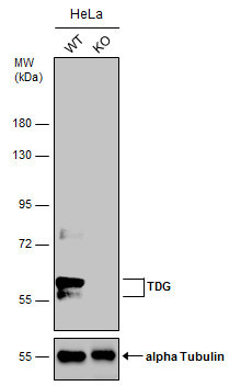
- Wild-type (WT) and TDG knockout (KO) HeLa cell extracts (30 ?g) were separated by 7.5% SDS-PAGE, and the membrane was blotted with TDG antibody (GTX110473) diluted at 1:500. The HRP-conjugated anti-rabbit IgG antibody (GTX213110-01) was used to detect the primary antibody.
- Submitted by
- GeneTex (provider)
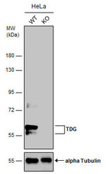
- Main image

- Experimental details
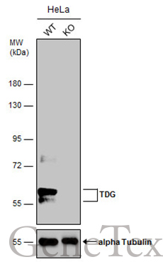
- Wild-type (WT) and TDG knockout (KO) HeLa cell extracts (30 ?g) were separated by 7.5% SDS-PAGE, and the membrane was blotted with TDG antibody (GTX110473) diluted at 1:500. The HRP-conjugated anti-rabbit IgG antibody (GTX213110-01) was used to detect the primary antibody.
Supportive validation
- Submitted by
- GeneTex (provider)
- Main image
- Experimental details
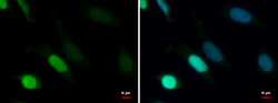
- TDG antibody detects TDG protein at nucleus by immunofluorescent analysis.Sample: HeLa cells were fixed in 4% paraformaldehyde at RT for 15 min.Green: TDG protein stained by TDG antibody (GTX110473) diluted at 1:1000.Blue: Hoechst 33342 staining.
