Explore
 Validate
 Learn
Learn Western blot
Antibody data
- Antibody Data
- Antigen structure
- References [1]
- Comments [0]
- Validations
- Western blot [4]
- Immunocytochemistry [1]
- Immunohistochemistry [2]
- Other assay [3]
Submit
Validation data
Reference
Comment
Report error
- Product number
- PA5-30237 - Provider product page

- Provider
- Invitrogen Antibodies
- Product name
- ERF Polyclonal Antibody
- Antibody type
- Polyclonal
- Antigen
- Recombinant protein fragment
- Description
- Recommended positive controls: A431, ERF-transfected 293T.
- Concentration
- 0.24 mg/mL
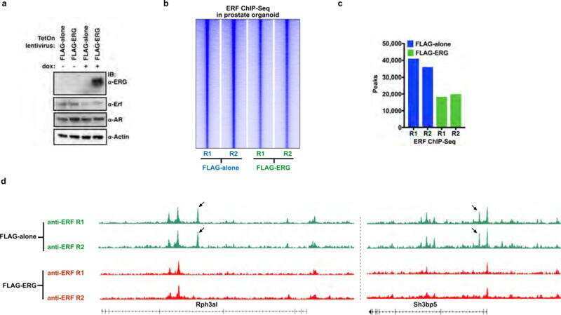
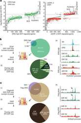
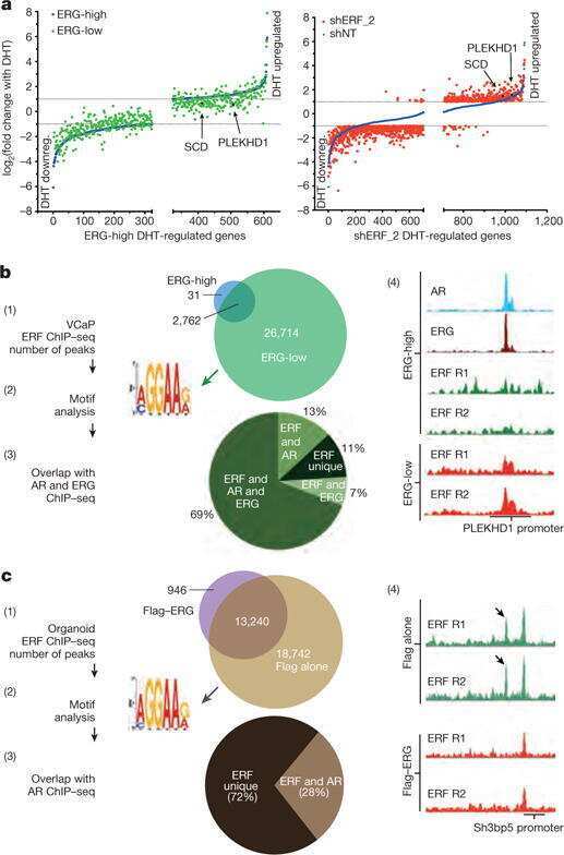
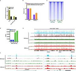
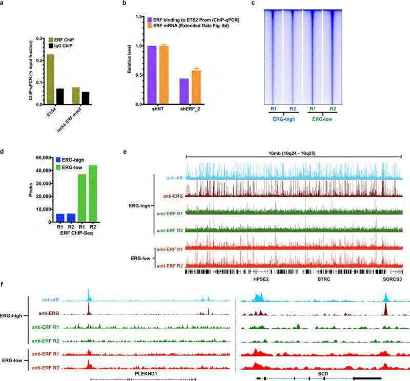
Submitted references ERF mutations reveal a balance of ETS factors controlling prostate oncogenesis.
Bose R, Karthaus WR, Armenia J, Abida W, Iaquinta PJ, Zhang Z, Wongvipat J, Wasmuth EV, Shah N, Sullivan PS, Doran MG, Wang P, Patruno A, Zhao Y, International SU2C/PCF Prostate Cancer Dream Team, Zheng D, Schultz N, Sawyers CL
Nature 2017 Jun 29;546(7660):671-675
Nature 2017 Jun 29;546(7660):671-675
No comments: Submit comment
Supportive validation
- Submitted by
- Invitrogen Antibodies (provider)
- Main image
- Experimental details
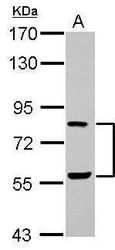
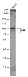
- Western blot analysis of ERF using 30 µg of A431 lysate. Samples were loaded onto a 7.5% SDS-PAGE gel and probed with an ERF polyclonal antibody (Product # PA5-30237) at a dilution of 1:1000.

- Submitted by
- Invitrogen Antibodies (provider)
- Main image

- Experimental details
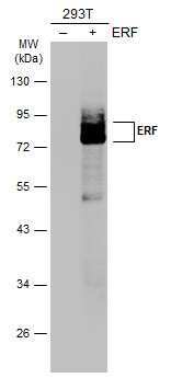
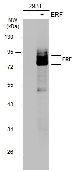
- Western Blot analysis of ERF was performed by separating 30 µg of non-transfected (–) and transfected (+) 293T whole cell extracts by 10% SDS-PAGE. Proteins were transferred to a membrane and probed with a ERF Polyclonal Antibody (Product # PA5-30237) at a dilution of 1:4000. The HRP-conjugated anti-rabbit IgG antibody was used to detect the primary antibody.
- Submitted by
- Invitrogen Antibodies (provider)
- Main image
- Experimental details
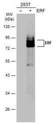
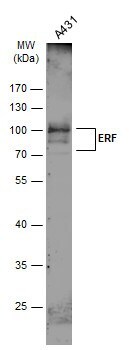
- Western blot analysis of ERF was performed by separating 30 µg of whole cell extract by 10% SDS-PAGE. Proteins were transferred to a membrane and probed with a ERF Polyclonal Antibody (Product # PA5-30237) at a dilution of 1:1000. The HRP-conjugated anti-rabbit IgG antibody was used to detect the primary antibody.
Supportive validation
- Submitted by
- Invitrogen Antibodies (provider)
- Main image
- Experimental details
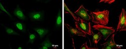
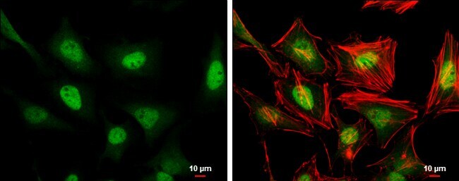
- Immunocytochemistry-Immunofluorescence analysis of ERF was performed in HeLa cells fixed in 4% paraformaldehyde at RT for 15 min. Green: ERF Polyclonal Antibody (Product # PA5-30237) diluted at 1:500. Red: phalloidin, a cytoskeleton marker. Scale bar = 10 µm.
Supportive validation
- Submitted by
- Invitrogen Antibodies (provider)
- Main image

- Experimental details
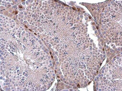
- Immunohistochemistry (Paraffin) analysis of ERF was performed in paraffin-embedded mouse testis tissue using ERF Polyclonal Antibody (Product # PA5-30237) at a dilution of 1:500.
- Submitted by
- Invitrogen Antibodies (provider)
- Main image
- Experimental details
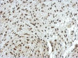
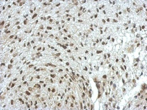
- Immunohistochemical analysis of paraffin-embedded U373 xenograft, using ERF (Product # PA5-30237) antibody at 1:500 dilution. Antigen Retrieval: EDTA based buffer, pH 8.0, 15 min.
Supportive validation
- Submitted by
- Invitrogen Antibodies (provider)
- Main image
- Experimental details
- NULL
- Submitted by
- Invitrogen Antibodies (provider)
- Main image
- Experimental details
- NULL
- Submitted by
- Invitrogen Antibodies (provider)
- Main image
- Experimental details
- NULL