Explore
 Validate
 Learn
Learn Western blot
Antibody data
- Antibody Data
- Antigen structure
- References [1]
- Comments [0]
- Validations
- Western blot [4]
- Immunocytochemistry [1]
- Immunohistochemistry [2]
Submit
Validation data
Reference
Comment
Report error
- Product number
- PA5-81235 - Provider product page

- Provider
- Invitrogen Antibodies
- Product name
- PAX2 Polyclonal Antibody
- Antibody type
- Polyclonal
- Antigen
- Synthetic peptide
- Description
- This product is preservative free. It is recommended to add sodium azide to avoid contamination (final concentration 0.05%-0.1%).
- Concentration
- 1 mg/mL
Submitted references Derivation of dorsal spinal sensory interneurons from human pluripotent stem cells.
Gupta S, Yamauchi K, Novitch BG, Butler SJ
STAR protocols 2021 Mar 19;2(1):100319
STAR protocols 2021 Mar 19;2(1):100319
No comments: Submit comment
Supportive validation
- Submitted by
- Invitrogen Antibodies (provider)
- Main image

- Experimental details
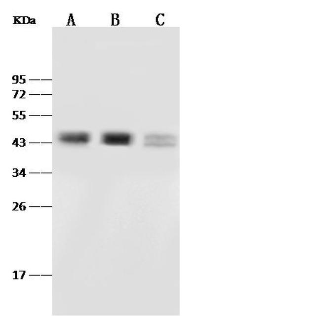
- Western blot analysis of PAX2 in Lane A: K562 Whole Cell Lysate (30 µg), Lane B: 293T Whole Cell Lysate (30 µg), Lane C: MCF7 Whole Cell Lysate (30 µg). Samples were probed using a PAX2 Polyclonal Antibody (Product # PA5-81235) at a 1:500 dilution, followed by a Goat Anti-Rabbit IgG (H+L), HRP Secondary Antibody at a 1:10000 dilution. Western blot was performed under reducing conditions. Predicted band size:44 kDa. Observed band size:44 kDa.
- Submitted by
- Invitrogen Antibodies (provider)
- Main image

- Experimental details
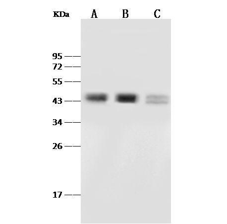
- Western Blot using PAX2 Polyclonal Antibody (Product # PA5-81235) at 1:500 dilution. Lane A: K562 Whole Cell Lysate, Lane B: 293T Whole Cell Lysate, Lane C: MCF7 Whole Cell Lysate. Lysates/proteins at 30 μg per lane. Secondary antibody: Goat Anti-Rabbit IgG (H+L)/HRP at 1:10,000 dilution. Developed using the ECL technique. Performed under reducing conditions. Predicted band size: 44 kDa. Observed band size: 44 kDa.
- Submitted by
- Invitrogen Antibodies (provider)
- Main image
- Experimental details
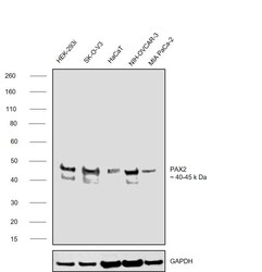
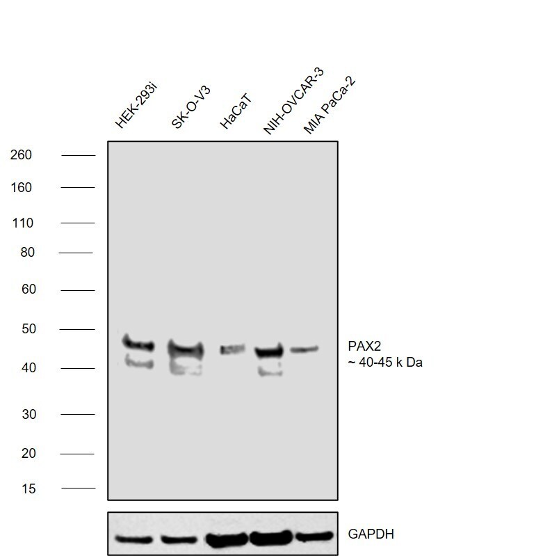
- Western blot was performed using Anti-PAX2 Polyclonal Antibody (Product # PA5-81235) and a 40 to 45 kDa band corresponding to Paired box protein Pax-2 was observed across cell lines tested . Nuclear enriched extracts (50 µg lysate) of HEK-293 (Lane 1), SK-O-V3 (Lane 2), HaCaT (Lane 3), NIH:OVCAR-3 (Lane 4), MIA PaCa-2 (Lane 5) were electrophoresed using NuPAGE™ 4-12% Bis-Tris Protein Gel (Product # NP0321BOX). Resolved proteins were then transferred onto a nitrocellulose membrane (Product # IB23001) by iBlot® 2 Dry Blotting System (Product # IB21001). The blot was probed with the primary antibody (1:1000 dilution) and detected by chemiluminescence with Goat anti-Rabbit IgG (H+L) Superclonal™ Recombinant Secondary Antibody, HRP (Product # A27036,1:4000) using the iBright FL 1000 (Product # A32752). Chemiluminescent detection was performed using SuperSignal™ West Pico PLUS Chemiluminescent Substrate (Product # 34580).
- Submitted by
- Invitrogen Antibodies (provider)
- Main image
- Experimental details
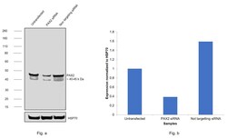
- Knockdown of Paired box protein Pax-2 was achieved by transfecting HEK-293 with Paired box protein Pax-2 specific siRNAs (Silencer® select Product # S532092, S532093). Western blot analysis (Fig. a) was performed using Nuclear enriched extracts from the Paired box protein Pax-2 knockdown cells (lane 2), non-targeting scrambled siRNA transfected cells (lane 3) and untransfected cells (lane 1). The blot was probed with PAX2 Polyclonal Antibody (Product # PA5-81235, 1:4000 ) and Goat anti-Rabbit IgG (H+L) Superclonal™ Recombinant Secondary Antibody, HRP (Product # A27036, 1:4000). Densitometric analysis of this western blot is shown in histogram (Fig. b). Decrease in signal upon siRNA mediated knock down confirms that antibody is specific to Paired box protein Pax-2.

Supportive validation
- Submitted by
- Invitrogen Antibodies (provider)
- Main image
- Experimental details
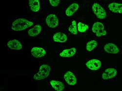
- Immunofluorescence staining of PAX2 in A549 cells. Cells were fixed with 4% PFA, permeabilzed with 0.1% Triton X-100 in PBS, blocked with 10% serum, and incubated with PAX2 Polyclonal Antibody (Product # PA5-81235, 1:1,000) at 4°C overnight. Then cells were stained with the Alexa Fluor®488-conjugated Goat Anti-rabbit IgG secondary antibody (green). Positive staining was localized to nucleus.

Supportive validation
- Submitted by
- Invitrogen Antibodies (provider)
- Main image
- Experimental details
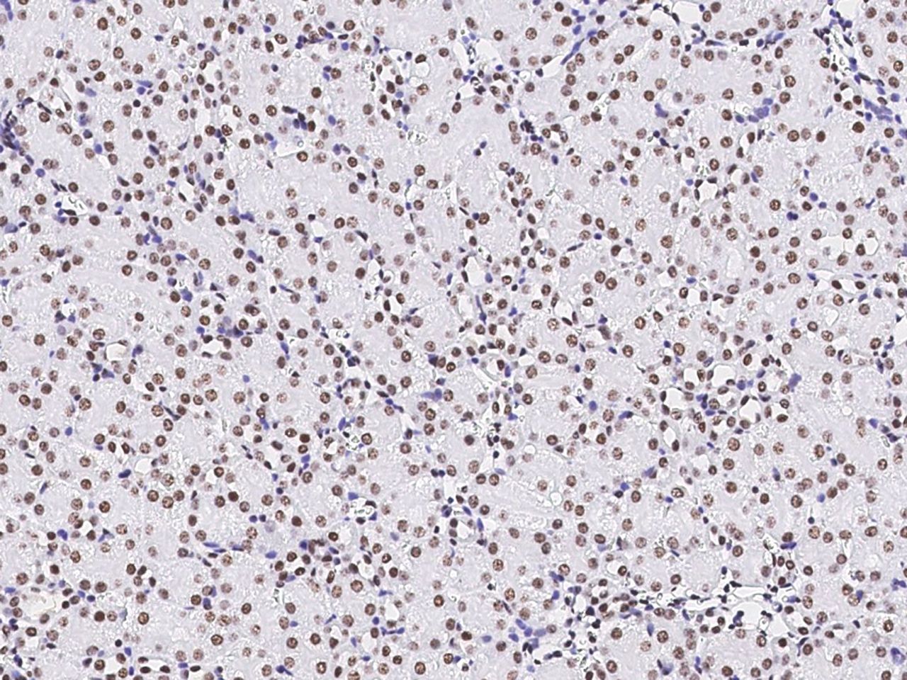
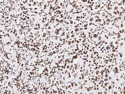
- Immunohistochemical staining of PAX2 in human kidney with PAX2 Polyclonal Antibody (Product # PA5-81235, 1:5,000, formalin-fixed paraffin embedded sections).
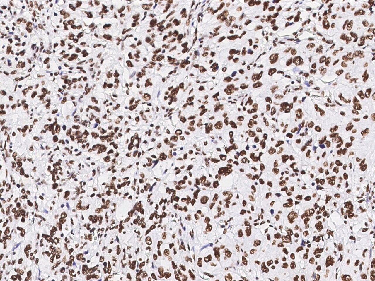
- Submitted by
- Invitrogen Antibodies (provider)
- Main image
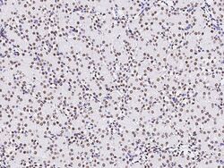
- Experimental details
- Immunohistochemical staining of PAX2 in mouse kidney with PAX2 Polyclonal Antibody (Product # PA5-81235, 1:5,000, formalin-fixed paraffin embedded sections).