Explore
 Validate
 Learn
Learn Western blot
Antibody data
- Antibody Data
- Antigen structure
- References [0]
- Comments [0]
- Validations
- Western blot [2]
- Immunocytochemistry [2]
- Immunohistochemistry [1]
Submit
Validation data
Reference
Comment
Report error
- Product number
- PA5-59539 - Provider product page

- Provider
- Invitrogen Antibodies
- Product name
- OAS3 Polyclonal Antibody
- Antibody type
- Polyclonal
- Antigen
- Recombinant full-length protein
- Description
- Immunogen sequence: TWDLGNGAAW HWDLLAQEAA SCYDHPCFLR GMGDPVQSWK GPGLPRAGCS GLGHPIQLDP NQKTPENSKS LNAVYPRAGS KPPSCP
- Concentration
- 0.4 mg/mL
No comments: Submit comment
Supportive validation
- Submitted by
- Invitrogen Antibodies (provider)
- Main image
- Experimental details
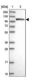
- Western blot analysis of OAS3 in Lane 1: Marker (kDa) 250, 130, 95, 72, 55, 36, 28, 17, 10; Lane 2: Human cell line RT-4. Samples were probed using an OAS3 Polyclonal Antibody (Product # PA5-59539).

- Submitted by
- Invitrogen Antibodies (provider)
- Main image
- Experimental details
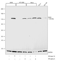
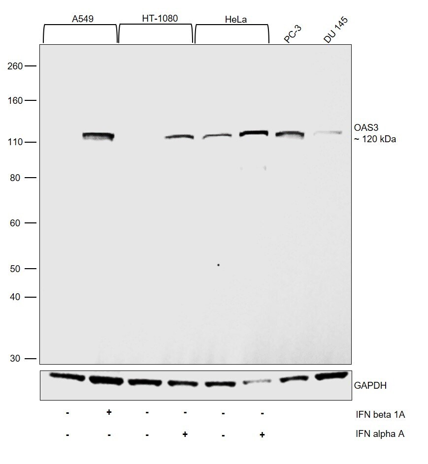
- Western Blot was performed using Anti-OAS3 Polyclonal Antibody (Product # PA5-59539) and a 120 kDa band corresponding to 2-5-oligoadenylate synthase 3 was observed. Whole cell extracts (30 µg lysate) of A549 (Lane 1), A549 treated with IFN beta 1A (1000 U/mL, O/N) (Lane 2), HT-1080 (Lane 3), HT-1080 treated with IFN alpha A (2000 U/mL, O/N) (Lane 4), HeLa (Lane 5), HeLa treated with IFN alpha A (10 ng/mL,16 hrs) (Lane 6), PC-3 (Lane 7) and DU 145 (Lane 8) were electrophoresed using NuPAGE™ 4-12% Bis-Tris Protein Gel (Product # NP0322BOX). Resolved proteins were then transferred onto a nitrocellulose membrane (Product # IB23001) by iBlot® 2 Dry Blotting System (Product # IB21001). The blot was probed with the primary antibody (0.2 µg/mL) and detected by chemiluminescence with Goat anti-Rabbit IgG (H+L) Superclonal™ Recombinant Secondary Antibody, HRP (Product # A27036, 1:4000) using the iBright FL 1000 (Product # A32752). Chemiluminescent detection was performed using Novex® ECL Chemiluminescent Substrate Reagent Kit (Product # WP20005). Upregulation in the expression of OAS3 was observed upon Interferon treatment in all the models tested (https://doi.org/10.1073/pnas.1519657113).
Supportive validation
- Submitted by
- Invitrogen Antibodies (provider)
- Main image
- Experimental details
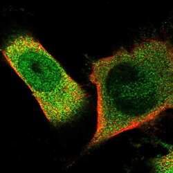
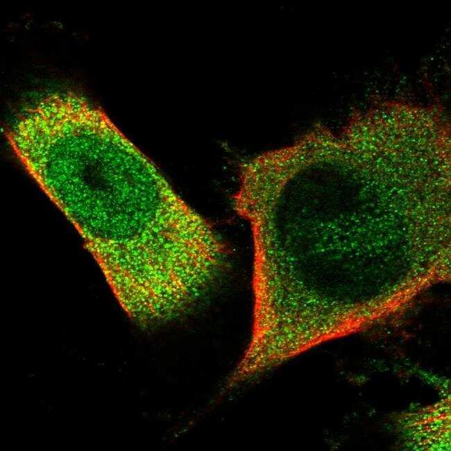
- Immunofluorescent staining of OAS3 in human cell line U-251 MG shows positivity in cytoplasm & nucleus but excluded from the nucleoli. Samples were probed using an OAS3 Polyclonal Antibody (Product # PA5-59539).
- Submitted by
- Invitrogen Antibodies (provider)
- Main image
- Experimental details
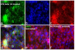
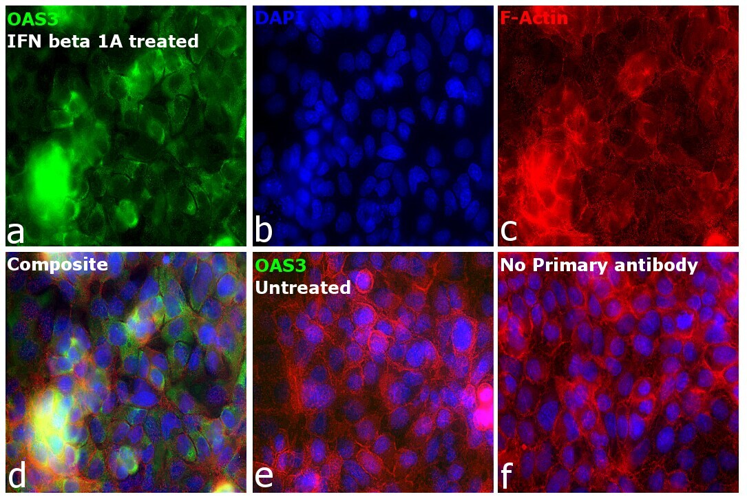
- Immunofluorescence analysis of 2-5-oligoadenylate synthase 3 was performed using 70% confluent log phase A549 treated with IFN beta 1A (1000 U/mL, O/N). The cells were fixed with 4% paraformaldehyde for 10 minutes, permeabilized with 0.1% Triton™ X-100 for 15 minutes, and blocked with 2% BSA for 1 hour at room temperature. The cells were labeled with OAS3 Polyclonal Antibody (Product # PA5-59539) at 1 µg/mL in 0.1% BSA, incubated at 4 degree celsius overnight and then labeled with Donkey anti-Rabbit IgG (H+L) Highly Cross-Adsorbed Secondary Antibody, Alexa Fluor Plus 488 (Product # A32790), (1:2000), for 45 minutes at room temperature (Panel a: Green). Nuclei (Panel b:Blue) were stained with ProLong™ Diamond Antifade Mountant with DAPI (Product # P36962). F-actin (Panel c: Red) was stained with Rhodamine Phalloidin (Product # R415, 1:300). Panel d represents the merged image showing predominant cytoplasmic localization. Panel e represents untreated A549 cells. Panel f represents control cells with no primary antibody to assess background. The images were captured at 60X magnification in EVOS™ M7000 Imaging System (Product # AMF7000).
Supportive validation
- Submitted by
- Invitrogen Antibodies (provider)
- Main image

- Experimental details
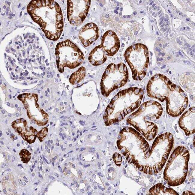
- Immunohistochemical staining of OAS3 in human kidney tissue shows strong cytoplasmic and membranous positivity in cells in tubules. Samples were probed using an OAS3 Polyclonal Antibody (Product # PA5-59539).