 Explore
 Validate
 Learn
Learn Western blot
 Immunocytochemistry
Antibody data
- Antibody Data
- Antigen structure
- References [8]
- Comments [0]
- Validations
- Immunocytochemistry [1]
- Immunohistochemistry [1]
Submit
Validation data
Reference
Comment
Report error
- Product number
- AF719 - Provider product page

- Provider
- R&D Systems
- Product name
- Mouse Noggin Antibody
- Antibody type
- Polyclonal
- Description
- Immunogen affinity purified. Detects mouse Noggin in direct ELISAs and Western blots. In direct ELISAs, approximately 30% cross-reactivity with recombinant human Noggin is observed.
- Reactivity
- Mouse
- Host
- Goat
- Conjugate
- Unconjugated
- Antigen sequence
P97466- Isotype
- IgG
- Vial size
- 100 ug
- Concentration
- LYOPH
- Storage
- Use a manual defrost freezer and avoid repeated freeze-thaw cycles. 12 months from date of receipt, -20 to -70 °C as supplied. 1 month, 2 to 8 °C under sterile conditions after reconstitution. 6 months, -20 to -70 °C under sterile conditions after reconstitution.
Submitted references Rhabdomyosarcoma and Wilms tumors contain a subpopulation of noggin producing, myogenic cells immunoreactive for lens beaded filament proteins.
Noggin depletion in adipocytes promotes obesity in mice.
Myo/Nog cells are present in the ciliary processes, on the zonule of Zinn and posterior capsule of the lens following cataract surgery.
Myo/Nog cells: targets for preventing the accumulation of skeletal muscle-like cells in the human lens.
Joint TGF-β type II receptor-expressing cells: ontogeny and characterization as joint progenitors.
BMP signalling permits population expansion by preventing premature myogenic differentiation in muscle satellite cells.
Noggin producing, MyoD-positive cells are crucial for eye development.
MyoD-positive epiblast cells regulate skeletal muscle differentiation in the embryo.
Gerhart J, Behling K, Paessler M, Milton L, Bramblett G, Garcia D, Pitts M, Hurtt R, Crawford M, Lackman R, Nguyen D, Infanti J, FitzGerald P, George-Weinstein M
PloS one 2019;14(4):e0214758
PloS one 2019;14(4):e0214758
Noggin depletion in adipocytes promotes obesity in mice.
Blázquez-Medela AM, Jumabay M, Rajbhandari P, Sallam T, Guo Y, Yao J, Vergnes L, Reue K, Zhang L, Yao Y, Fogelman AM, Tontonoz P, Lusis AJ, Wu X, Boström KI
Molecular metabolism 2019 Jul;25:50-63
Molecular metabolism 2019 Jul;25:50-63
Myo/Nog cells are present in the ciliary processes, on the zonule of Zinn and posterior capsule of the lens following cataract surgery.
Gerhart J, Withers C, Gerhart C, Werner L, Mamalis N, Bravo-Nuevo A, Scheinfeld V, FitzGerald P, Getts R, George-Weinstein M
Experimental eye research 2018 Jun;171:101-105
Experimental eye research 2018 Jun;171:101-105
Myo/Nog cells: targets for preventing the accumulation of skeletal muscle-like cells in the human lens.
Gerhart J, Greenbaum M, Scheinfeld V, Fitzgerald P, Crawford M, Bravo-Nuevo A, Pitts M, George-Weinstein M
PloS one 2014;9(4):e95262
PloS one 2014;9(4):e95262
Joint TGF-β type II receptor-expressing cells: ontogeny and characterization as joint progenitors.
Li T, Longobardi L, Myers TJ, Temple JD, Chandler RL, Ozkan H, Contaldo C, Spagnoli A
Stem cells and development 2013 May 1;22(9):1342-59
Stem cells and development 2013 May 1;22(9):1342-59
BMP signalling permits population expansion by preventing premature myogenic differentiation in muscle satellite cells.
Ono Y, Calhabeu F, Morgan JE, Katagiri T, Amthor H, Zammit PS
Cell death and differentiation 2011 Feb;18(2):222-34
Cell death and differentiation 2011 Feb;18(2):222-34
Noggin producing, MyoD-positive cells are crucial for eye development.
Gerhart J, Pfautz J, Neely C, Elder J, DuPrey K, Menko AS, Knudsen K, George-Weinstein M
Developmental biology 2009 Dec 1;336(1):30-41
Developmental biology 2009 Dec 1;336(1):30-41
MyoD-positive epiblast cells regulate skeletal muscle differentiation in the embryo.
Gerhart J, Elder J, Neely C, Schure J, Kvist T, Knudsen K, George-Weinstein M
The Journal of cell biology 2006 Oct 23;175(2):283-92
The Journal of cell biology 2006 Oct 23;175(2):283-92
No comments: Submit comment
Supportive validation
- Submitted by
- R&D Systems (provider)
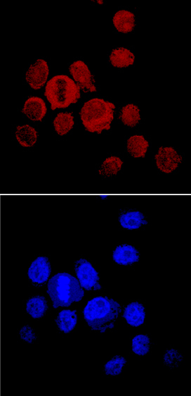
- Main image

- Experimental details
- Noggin in PC-3 Human Cell Line. Noggin was detected in immersion fixed PC-3 human prostate cancer cell line using Goat Anti-Mouse Noggin Antigen Affinity-purified Polyclonal Antibody (Catalog # AF719) at 10 µg/mL for 3 hours at room temperature. Cells were stained using the NorthernLights™ 557-conjugated Anti-Goat IgG Secondary Antibody (red, upper panel; Catalog # NL001) and counterstained with DAPI (blue, lower panel). Specific staining was localized to cytoplasm. View our protocol for Fluorescent ICC Staining of Cells on Coverslips.
Supportive validation
- Submitted by
- R&D Systems (provider)
- Main image
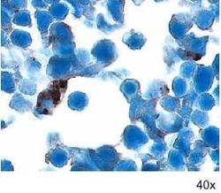
- Experimental details
- Noggin in Embryonic Mouse Cardiac Tissue. Noggin was detected in immersion fixed frozen sections of embryonic mouse cardiac tissue (11 d.p.c.) using 15 µg/mL Goat Anti-Mouse Noggin Antigen Affinity-purified Polyclonal Antibody (Catalog # AF719) overnight at 4 °C. Tissue was stained with the Anti-Goat HRP-DAB Cell & Tissue Staining Kit (brown; Catalog # CTS008) and counterstained with hematoxylin (blue). View our protocol for Chromogenic IHC Staining of Frozen Tissue Sections.
