Explore
 Validate
 Learn
Learn Western blot
Antibody data
- Antibody Data
- Antigen structure
- References [1]
- Comments [0]
- Validations
- Western blot [3]
- Immunocytochemistry [1]
Submit
Validation data
Reference
Comment
Report error
- Product number
- MA1-19373 - Provider product page

- Provider
- Invitrogen Antibodies
- Product name
- SOCS3 Monoclonal Antibody (SO1)
- Antibody type
- Monoclonal
- Antigen
- Other
- Description
- Western Blot: Reducing conditions.
- Reactivity
- Human
- Host
- Mouse
- Isotype
- IgG
- Antibody clone number
- SO1
- Vial size
- 100 µg
- Concentration
- 1 mg/mL
- Storage
- 4° C, do not freeze
Submitted references Chemokine CCL5 promotes robust optic nerve regeneration and mediates many of the effects of CNTF gene therapy.
Xie L, Yin Y, Benowitz L
Proceedings of the National Academy of Sciences of the United States of America 2021 Mar 2;118(9)
Proceedings of the National Academy of Sciences of the United States of America 2021 Mar 2;118(9)
No comments: Submit comment
Supportive validation
- Submitted by
- Invitrogen Antibodies (provider)
- Main image

- Experimental details
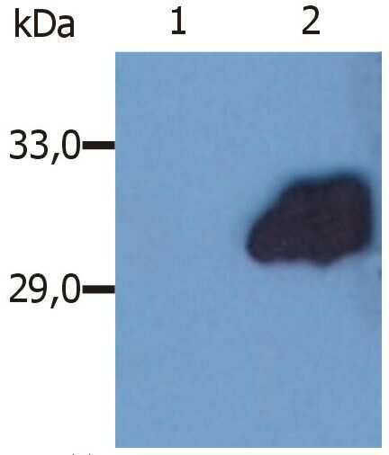
- Western blotting analysis (reducing conditions) of whole cell lysate of HeLa human cervix carcinoma cell line. Lane 1: immunostaining with Isotype mouse IgG2b control (PLRV219); Lane 2: immunostaining with anti-SOCS3 (SO1) Monoclonal antibody (Product # MA1-19373).
- Submitted by
- Invitrogen Antibodies (provider)
- Main image

- Experimental details
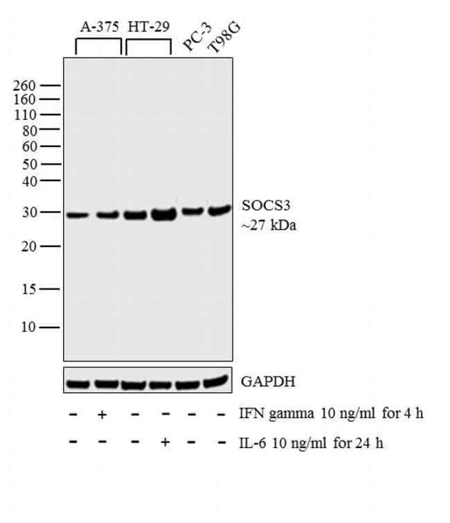
- Western blot analysis was performed on whole cell extracts (30 µg lysate) of A-375 (Lane 1), A-375 treated with IFN gamma (10 ng/mL for 4 h) (Lane 2), HT-29 (Lane 3), HT-29 treated with IL-6 (10 ng/mL for 24 h) (Lane 4), PC-3 (Lane 5) and T98G (Lane 6). The blot was probed with Anti-SOCS3 Monoclonal Antibody (Product # MA1-19373, 1 µg/mL) and detected by chemiluminescence using Goat anti-Mouse IgG (H+L) Superclonal™ Secondary Antibody, HRP conjugate (Product # A28177, 0.25 µg/mL, 1:4000 dilution). A 27 kDa band corresponding to SOCS3 was observed across the cell lines tested and was enhanced upon treatment.
- Submitted by
- Invitrogen Antibodies (provider)
- Main image

- Experimental details
- Western blotting analysis (reducing conditions) of whole cell lysate of HeLa human cervix carcinoma cell line. Lane 1: immunostaining with Isotype mouse IgG2b control (PLRV219); Lane 2: immunostaining with anti-SOCS3 (SO1) Monoclonal antibody (Product # MA1-19373).

Supportive validation
- Submitted by
- Invitrogen Antibodies (provider)
- Main image
- Experimental details
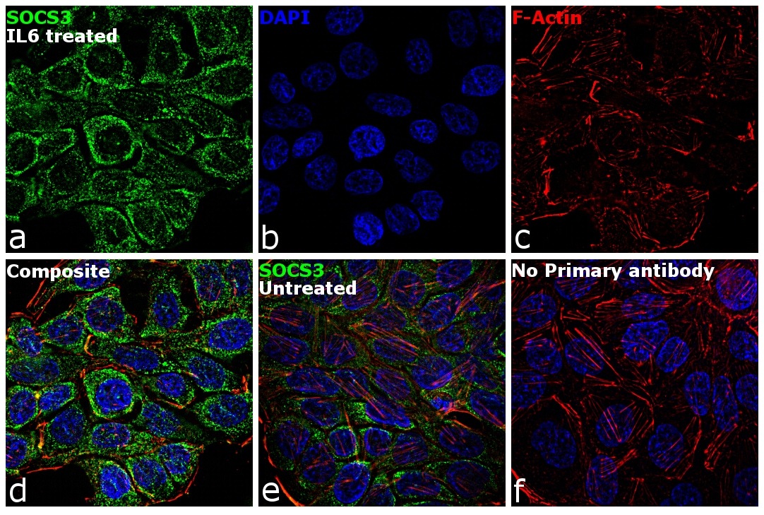
- Immunofluorescence analysis of SOCS3 was performed using 70% confluent log phase HT-29 cells treated with 10 ng of IL-6 for 24 hours. The cells were fixed with 4% paraformaldehyde for 10 minutes, permeabilized with 0.1% Triton™ X-100 for 15 minutes, and blocked with 1% BSA for 1 hour at room temperature. The cells were labeled with SOCS3 Mouse Monoclonal Antibody (Product # MA1-19373) at 5 µg/mL in 0.1% BSA, incubated at 4 degree Celsius overnight and then labeled with Goat anti-Mouse IgG (H+L) Superclonal™ Secondary Antibody, Alexa Fluor® 488 conjugate (Product # A28175) at a dilution of 1:2000 for 45 minutes at room temperature (Panel a: green). Nuclei (Panel b: blue) were stained with SlowFade® Gold Antifade Mountant with DAPI (Product # S36938). F-actin (Panel c: red) was stained with Rhodamine Phalloidin (Product # R415, 1:300). Panel d represents the merged image showing cytoplasmic localization. Panel e shows untreated cells with less cytoplasmic signal. Panel f represents control cells with no primary antibody to assess background. The images were captured at 60X magnification.