Explore
 Validate
 Learn
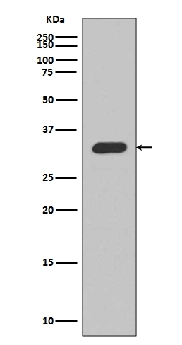
Learn Western blot
 Immunohistochemistry
Antibody data
- Antibody Data
- Antigen structure
- References [0]
- Comments [0]
- Validations
- Immunohistochemistry [1]
Submit
Validation data
Reference
Comment
Report error
- Product number
- M00173-1 - Provider product page

- Provider
- Boster Biological Technology
- Product name
- Anti-p27 KIP 1 CDKN1B Rabbit Monoclonal Antibody
- Antibody type
- Monoclonal
- Description
- Monoclonal antibody for p27/CDKN1B detection. Host: Rabbit.Size: 100ug/vial. Tested applications: Flow Cytometry, IP, IF, IHC, ICC, WB. Reactive species: Human, Rat p27/CDKN1B information: Molecular Weight: 22073 MW; Subcellular Localization: Nucleus. Cytoplasm. Endosome . Nuclear and cytoplasmic in quiescent cells. AKT- or RSK- mediated phosphorylation on Thr-198, binds 14-3-3, translocates to the cytoplasm and promotes cell cycle progression. Mitogen- activated UHMK1 phosphorylation on Ser-10 also results in translocation to the cytoplasm and cell cycle progression. Phosphorylation on Ser-10 facilitates nuclear export. Translocates to the nucleus on phosphorylation of Tyr-88 and Tyr-89. Colocalizes at the endosome with SNX6; this leads to lysosomal degradation (By similarity); Tissue Specificity: Expressed in all tissues tested. Highest levels in skeletal muscle, lowest in liver and kidney.
- Reactivity
- Human, Rat
- Host
- Rabbit
- Antibody clone number
- CGA-3
- Vial size
- 100ug/vial
- Concentration
- 0.5-1mg/ml, actual concentration vary by lot. Use suggested dilution ratio to decide dilution procedure.
- Storage
- At -20°C for one year. Avoid repeated freezing and thawing.
No comments: Submit comment
Supportive validation
- Submitted by
- Boster Biological Technology (provider)
- Main image
- Experimental details
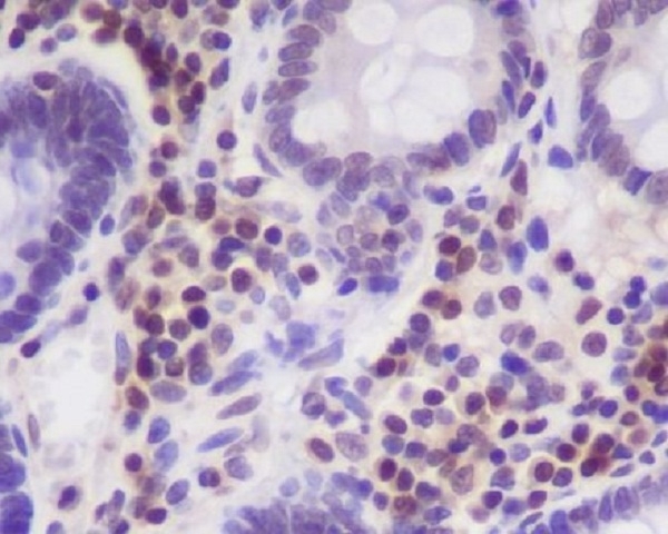
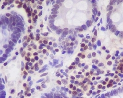
- Immunohistochemical analysis of paraffin-embedded human colon, using p27 KIP 1 Antibody(M00173-1)CDKN1B was detected in paraffin-embedded tissue section. Heat mediated antigen retrieval was performed in citrate buffer (pH6, epitope retrieval solution) for 20 mins. The tissue section was blocked with 10% goat serum. The tissue section was then incubated with 1ug/ml rabbit anti-CDKN1B Antibody (M00173-1)overnight at 4?? Biotinylated goat anti-rabbit IgG was used as secondary antibody and incubated for 30 minutes at 37?? The tissue section was developed using Strepavidin-Biotin-Complex (SABC)(Catalog # SA1022) with DAB as the chromogen.
- Additional image