Explore
 Validate
 Learn
Learn Western blot
Antibody data
- Antibody Data
- Antigen structure
- References [3]
- Comments [0]
- Validations
- Western blot [1]
- Immunohistochemistry [1]
- Other assay [8]
Submit
Validation data
Reference
Comment
Report error
- Product number
- PA5-43746 - Provider product page

- Provider
- Invitrogen Antibodies
- Product name
- CHI3L1 Polyclonal Antibody
- Antibody type
- Polyclonal
- Antigen
- Synthetic peptide
- Description
- Peptide sequence: LDFISIMTYD FHGAWRGTTG HHSPLFRGQE DASPDRFSNT DYAVGYMLRL
- Concentration
- 0.5 mg/mL
Submitted references YKL40 in sporadic amyotrophic lateral sclerosis: cerebrospinal fluid levels as a prognosis marker of disease progression.
Sporadic Creutzfeldt-Jakob disease with glial PrP(Res) nuclear and perinuclear immunoreactivity.
YKL-40 in the brain and cerebrospinal fluid of neurodegenerative dementias.
Andrés-Benito P, Domínguez R, Colomina MJ, Llorens F, Povedano M, Ferrer I
Aging 2018 Sep 13;10(9):2367-2382
Aging 2018 Sep 13;10(9):2367-2382
Sporadic Creutzfeldt-Jakob disease with glial PrP(Res) nuclear and perinuclear immunoreactivity.
Fernández-Vega I, Díaz-Lucena D, Azkune Calle I, Geijo M, Juste RA, Llorens F, Vicente Etxenausia I, Santos-Juanes J, Zarranz Imirizaldu JJ, Ferrer I
Neuropathology : official journal of the Japanese Society of Neuropathology 2018 Oct;38(5):561-567
Neuropathology : official journal of the Japanese Society of Neuropathology 2018 Oct;38(5):561-567
YKL-40 in the brain and cerebrospinal fluid of neurodegenerative dementias.
Llorens F, Thüne K, Tahir W, Kanata E, Diaz-Lucena D, Xanthopoulos K, Kovatsi E, Pleschka C, Garcia-Esparcia P, Schmitz M, Ozbay D, Correia S, Correia Â, Milosevic I, Andréoletti O, Fernández-Borges N, Vorberg IM, Glatzel M, Sklaviadis T, Torres JM, Krasemann S, Sánchez-Valle R, Ferrer I, Zerr I
Molecular neurodegeneration 2017 Nov 10;12(1):83
Molecular neurodegeneration 2017 Nov 10;12(1):83
No comments: Submit comment
Supportive validation
- Submitted by
- Invitrogen Antibodies (provider)
- Main image

- Experimental details
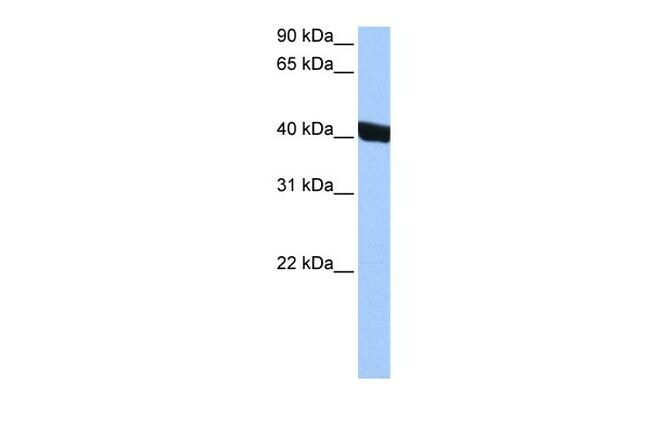
- Western blot analysis of human HepG2 cell lysate using an anti-CHI3L1 polyclonal antibody (Product # PA5-43746).
Supportive validation
- Submitted by
- Invitrogen Antibodies (provider)
- Main image
- Experimental details
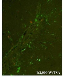
- Immunofluorescence analysis of Simian IVE tissue using an anti-CHI3L1 polyclonal antibody (Product # PA5-43746).

Supportive validation
- Submitted by
- Invitrogen Antibodies (provider)
- Main image
- Experimental details
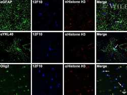
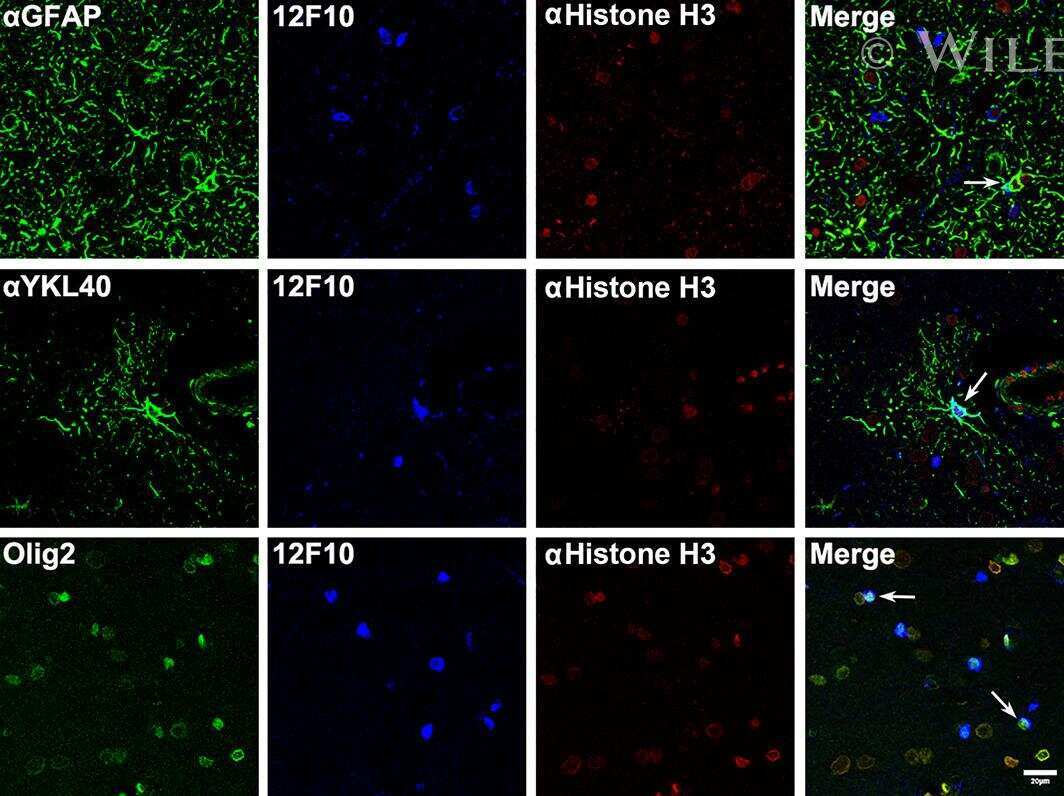
- Microphotographs by triple-labeling immunofluorescence and confocal microscopy using anti-GFAP, anti-YKL-40, Olig2, 12F10, and anti-histone H3. PrP immunoreactivity localizes in the nucleus of astrocytes and oligodendrocytes (arrows). Formalin-fixed, paraffin-embedded sections. Scale bars: 20 mum.
- Submitted by
- Invitrogen Antibodies (provider)
- Main image

- Experimental details
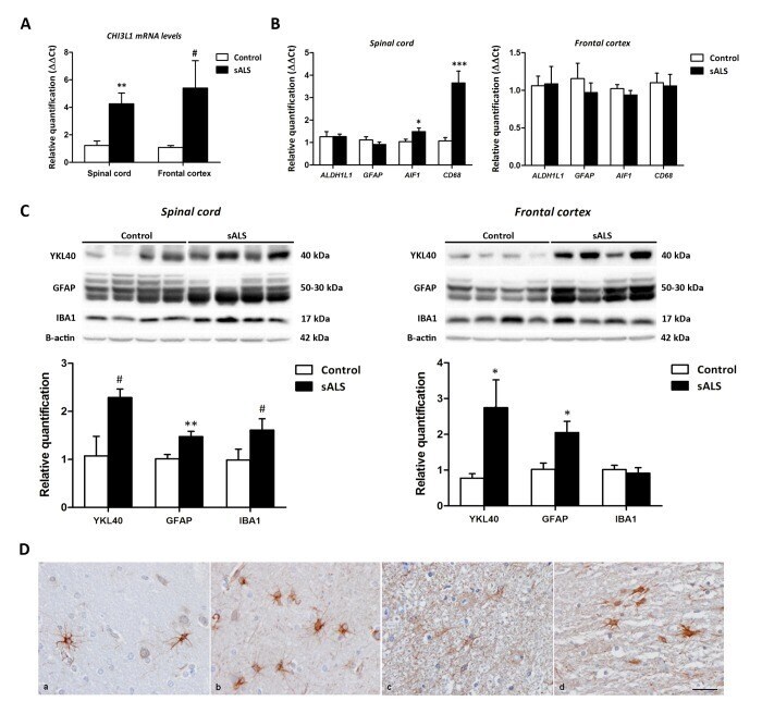
- Figure 1 ( A ) CHI3L1 mRNA expression levels in the anterior horn of the lumbar spinal cord and frontal cortex area 8 in sALS and control cases. CHI3L1 is significantly up-regulated in the anterior spinal cord but has only a tendency to increase without significance in the frontal cortex in sALS compared with controls. ( B ) mRNA expression levels of microglial ( CD68 and AIF1 ) and astroglial ( GFAP and ALDH1L1 ) markers in the anterior horn of lumbar spinal cord and frontal cortex area 8 in sALS and age-matched controls. Microglial markers CD68 and AIF1 are significantly up-regulated in the anterior horn of the spinal cord but not in the frontal cortex in sALS. The mRNA expression levels of astroglial markers in the spinal cord and frontal cortex are not modified in pathological cases when compared with controls. ( C ) Western blot analysis of YKL40 in the spinal cord (left panel) and frontal cortex area 8 (right panel) of control and sALS; beta-actin was used for normalization. Graphical representation of western blot data; fold changes in the expression of protein are determined relative to the control cases. YKL40 and GFAP protein levels are increased in the spinal cord and frontal cortex in sALS when compared with controls. Due to individual variation, increased values in the anterior horn of the spinal cord showed only a tendency without statistical significance. In contrast, expression levels were not significantly modified in sALS. * P < 0.05, ** P < 0.0
- Submitted by
- Invitrogen Antibodies (provider)
- Main image

- Experimental details
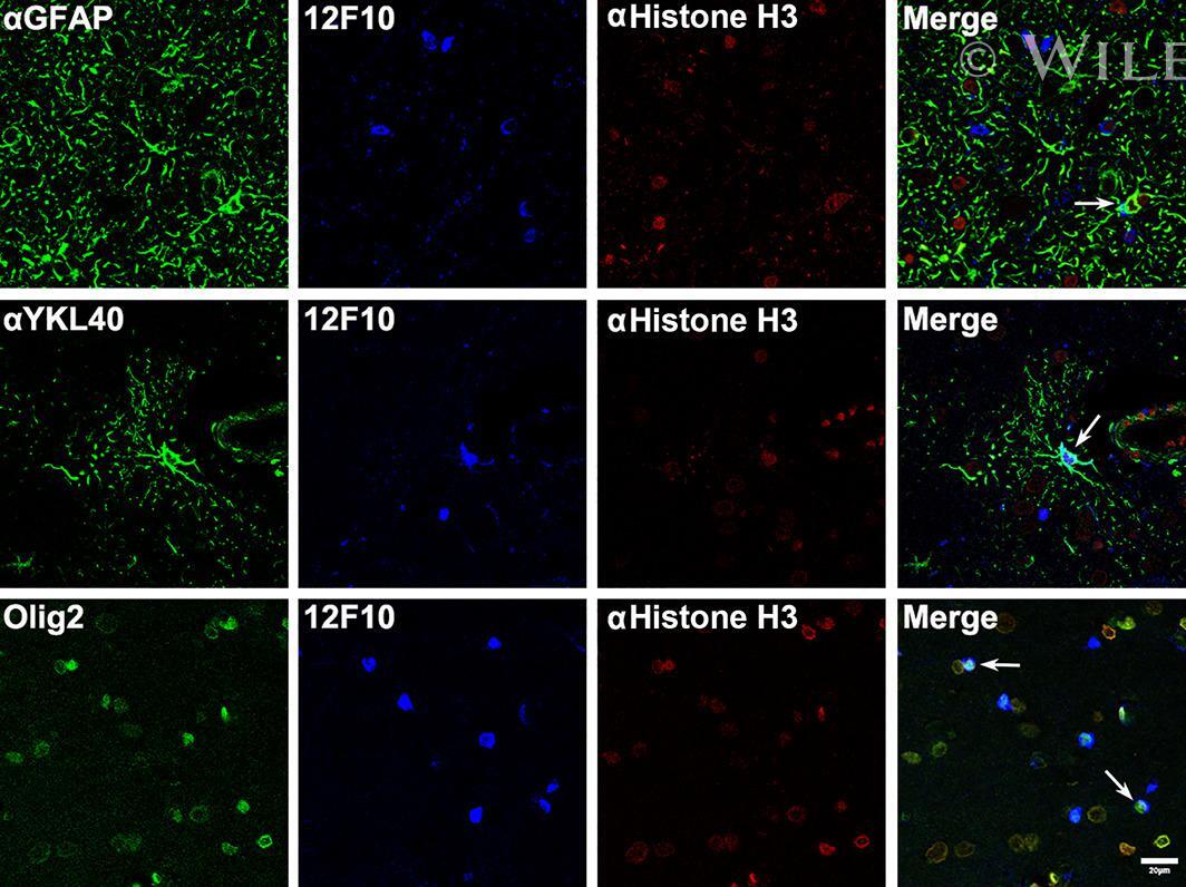
- Microphotographs by triple-labeling immunofluorescence and confocal microscopy using anti-GFAP, anti-YKL-40, Olig2, 12F10, and anti-histone H3. PrP immunoreactivity localizes in the nucleus of astrocytes and oligodendrocytes (arrows). Formalin-fixed, paraffin-embedded sections. Scale bars: 20 mum.
- Submitted by
- Invitrogen Antibodies (provider)
- Main image

- Experimental details
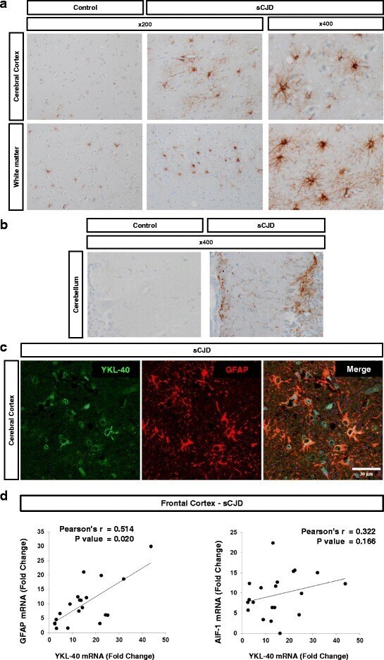
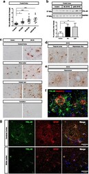
- Fig. 2 YKL-40 expression in sCJD brain tissue. a Immunohistochemical analysis for detection of YKL-40 in the cerebral cortex and white matter of control and sCJD cases. b Immunohistochemical analysis of YKL-40 in the cerebellum of control and sCJD cases. Brown staining corresponds to YKL-40 staining and light blue to haematoxylin counterstaining. c Immunofluorescence analysis of YKL-40 (green) and GFAP (red) in cerebral cortex region of sCJD. Nuclei were stained with DAPI (blue). d Correlation between the expression levels of YKL-40 and GFAP mRNA (left panel) or AIF-1 mRNA (right panel) in the frontal cortex of sCJD cases. Normalization was performed using GAPDH. Pearson test was used to determine the correlations between mRNA expression levels
- Submitted by
- Invitrogen Antibodies (provider)
- Main image
- Experimental details
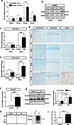
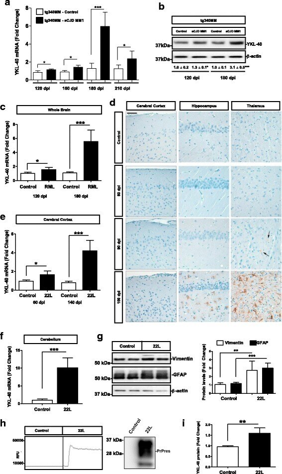
- Fig. 3 YKL-40 expression in experimental models of prion diseases. a RT-qPCR analysis of YKL-40 in the cortex of control and sCJD MM1 inoculated tg340 PRNP 129MM mice at 120 dpi (pre-clinical), 160 dpi (early clinical), 180 dpi (clinical) and 210 dpi (clinical with 10-1 diluted inoculum). Four animals per group were analyzed. Normalization was performed using Hprt. b Representative Western-blot analyses for YKL-40 immunodetection in the cortex of control and sCJD MM1-inoculated tg340 PRNP 129MM mice at 120 dpi (pre-clinical) and 180 dpi (clinical). Three animals per group were analyzed. Normalization was based on beta-actin levels. Numbers indicate densitometry results from three animals per group. Unpaired t-tests were performed to determine statistical differences. c RT-qPCR analysis of YKL-40 in the whole brain of control and RML scrapie-infected mice at pre-clinical (120 dpi) and clinical disease (180 dpi) stages. GAPDH was used for normalization. Similar results were acquired when normalization was based on Hprt expression levels (not shown). Unpaired t-tests were used for estimation of statistical differences. d Immunohistochemical analysis of YKL-40 expression in the cerebral cortex, hippocampus and thalamus of control and RML scrapie-infected mice at pre-clinical (60 and 90 dpi) and clinical (150 dpi) disease stages. Scale bar = 50 mum. Arrows indicate YKL-40 positive reactive astrocytes. Three animals per time point were used. Two sections were stained per animal (sa
- Submitted by
- Invitrogen Antibodies (provider)
- Main image
- Experimental details
- Fig. 4 YKL-40 expression in AD brain tissue ( a ) RT-qPCR analysis of YKL-40 in the frontal cortex of control, AD (I-III), AD (IV-VI) and rpAD (IV-VI) samples. GAPDH was used for normalization. Kruskal-Wallis and Dunn's post-hoc tests were used to estimate statistical differences. b Western blot analysis of YKL-40 in the frontal cortex of control, AD (IV-VI) and rpAD (IV-VI) samples. Normalization was based on GAPDH levels. Graphic summary of densitometry analyses performed on western blot results acquired from 8 control, 8 AD and 6 rpAD samples. c Immunohistochemical analysis of YKL-40 in the cerebral cortex, white matter, subpial layer and cerebellum in control and AD cases. d Immunohistochemical analysis of YKL-40 in the temporal cortex and hippocampus in AD cases. e Immunohistochemical analysis of YKL-40+ astrocytes surrounding beta-amyloid plaques (left) and in blood vessels with amyloid angiopathy (right) in the hippocampal region of AD cases Brown staining corresponds to YKL-40 staining and light blue to haematoxylin counterstaining. f Double-labeling immunofluorescence of YKL-40 (green) and amyloid beta (red) in the hippocampus of AD. g Double-labeling immunofluorescence of YKL-40 (green) and GFAP (red) in cerebral cortex and white matter in AD tissues. Fold changes in expression of mRNA and protein were determined relative to the control cases. * p < 0.05, *** p < 0.001

- Submitted by
- Invitrogen Antibodies (provider)
- Main image
- Experimental details
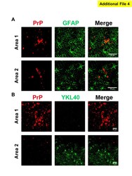
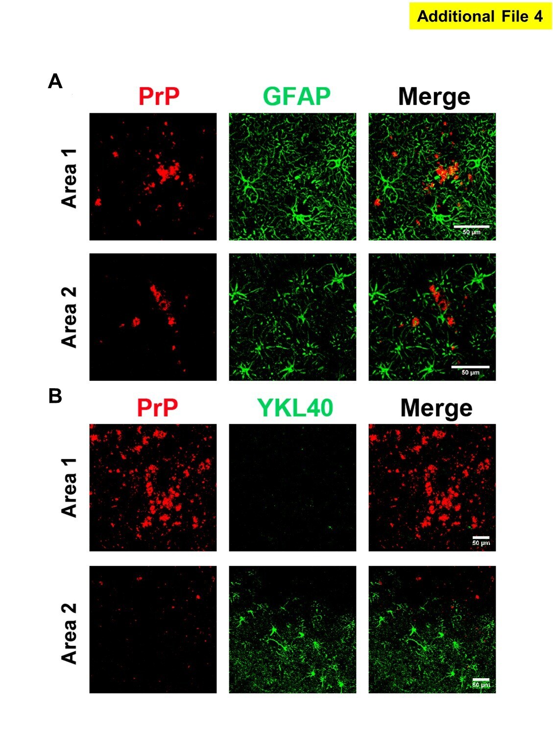
- Additional file 4: YKL-40-positive astrocytes associated to PrP amyloid plaques. Immunofluorescence images obtained from double-labeling staining with GFAP (A) and YKL-40 (B) (green) and PrP (red) antibodies in the cortex of sCJD. Individual channels as well as merge images are shown for two different cortical areas. (TIFF 768 kb)
- Submitted by
- Invitrogen Antibodies (provider)
- Main image
- Experimental details
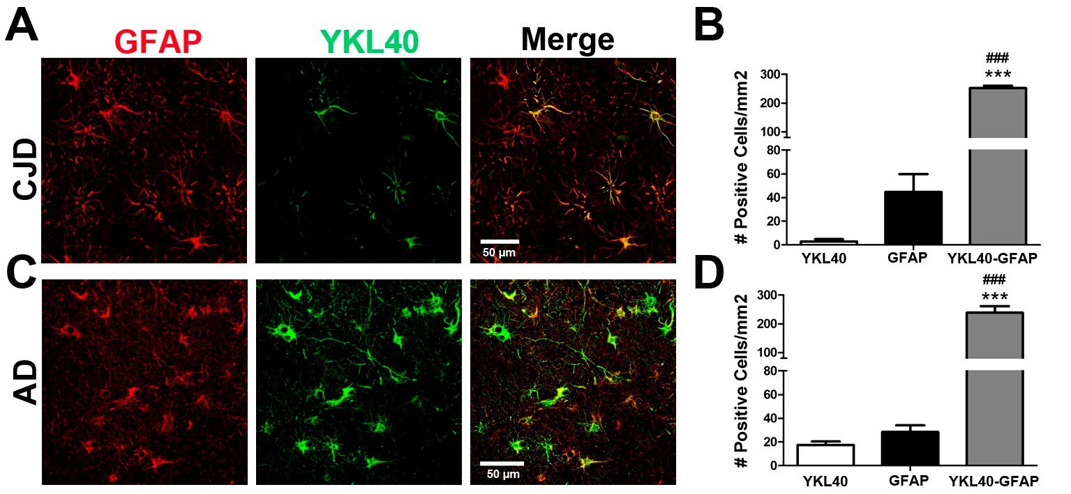
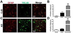
- Additional file 5: Quantification of YKL-40 and GFAP overlap in sCJD and AD cases. Immunofluorescence images obtained from double-labelling staining with GFAP (red) and YKL-40 (green) antibodies in the hippocampus of sCJD ( n = 3) (A) and AD V (n = 3) (C) cases. Individual channels as well as merge images are shown for two different cortical areas (one cortical image per CJD or AD patient) of two different patients. Quantifications of GFAP+/YKL-40+ (YKL-40-GFAP), GFAP+/YKL-40- (GFAP) and GFAP-/YKL-40+ (YKL-40) astrocytes are shown. Statistical significance differences were detected between YKL-40-GFAP and YKL-40, GFAP groups; ### p < 0.001 and *** p < 0.001, respectively. (TIFF 5257 kb)