Explore
 Validate
 Learn
Learn Western blot
Antibody data
- Antibody Data
- Antigen structure
- References [0]
- Comments [0]
- Validations
- Western blot [4]
- Immunohistochemistry [2]
Submit
Validation data
Reference
Comment
Report error
- Product number
- PA5-46996 - Provider product page

- Provider
- Invitrogen Antibodies
- Product name
- CHI3L1 Polyclonal Antibody
- Antibody type
- Polyclonal
- Antigen
- Recombinant full-length protein
- Description
- In direct ELISAs, approximately 15% cross-reactivity with recombinant mouse Chitinase 3-like 1 is observed.
- Concentration
- 0.2 mg/mL
No comments: Submit comment
Supportive validation
- Submitted by
- Invitrogen Antibodies (provider)
- Main image
- Experimental details
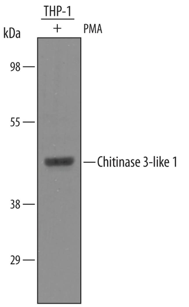
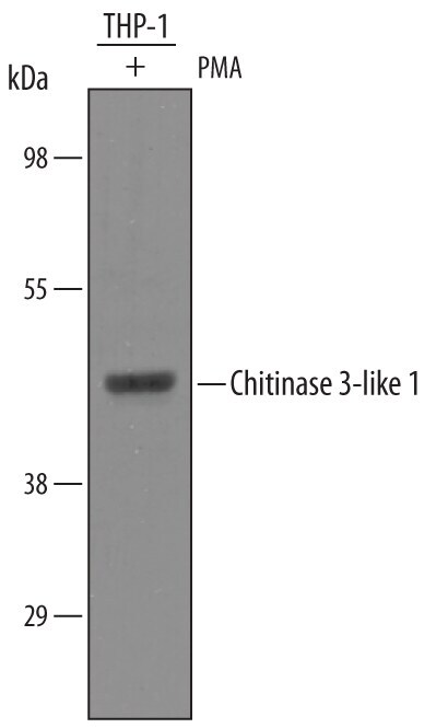
- Western blot analysis from lysates of THP-1 human acute monocytic leukemia cell line treated (+) with 50 ng/mL of PMA for 72 hours. PVDF membrane was probed with 1 µg/mL of Goat Anti-human Chitinase 3-like 1 Antigen Affinity-purified Polyclonal Antibody (Product # PA5-46996) followed by HRP-conjugated Anti-Goat IgG Secondary Antibody. A specific band was detected for Chitinase 3-like 1 at approximately 45 kDa (as indicated). This experiment was conducted under reducing conditions.

- Submitted by
- Invitrogen Antibodies (provider)
- Main image
- Experimental details
- Western blot analysis from lysates of THP-1 human acute monocytic leukemia cell line treated (+) with 50 ng/mL of PMA for 72 hours. PVDF membrane was probed with 1 µg/mL of Goat Anti-human Chitinase 3-like 1 Antigen Affinity-purified Polyclonal Antibody (Product # PA5-46996) followed by HRP-conjugated Anti-Goat IgG Secondary Antibody. A specific band was detected for Chitinase 3-like 1 at approximately 45 kDa (as indicated). This experiment was conducted under reducing conditions.
- Submitted by
- Invitrogen Antibodies (provider)
- Main image

- Experimental details
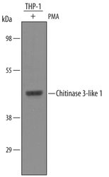
- Western blot analysis of CHI3L1 in THP‚1 human acute monocytic leukemia cell line treated (+) with 50 ng/mL of PMA for 72 hours. Samples were incubated in CHI3L1 polyclonal antibody (Product # PA5-46996) using a dilution of 1 µg/mL followed by a HRP-conjugated Anti-Goat IgG secondary antibody. A specific band was detected for Chitinase 3-like 1 at approximately 45 kDa (as indicated). This experiment was conducted under reducing conditions.
- Submitted by
- Invitrogen Antibodies (provider)
- Main image

- Experimental details
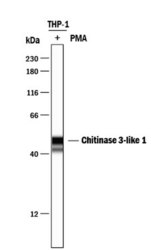
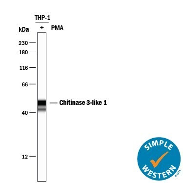
- Western blot analysis of CHI3L1 in 0.2 mg/mL lysates of THP‚1 human acute monocytic leukemia cell line treated (+) with 50 ng/mL PMA for 72 hours. Samples were incubated in CHI3L1 polyclonal antibody (Product # PA5-46996) using a dilution of 10 µg/mL followed by HRP-conjugated Anti-Goat IgG at a dilution of 0.0763888888888889. A specific band was detected for Chitinase 3‚like 1 at approximately 49 kDa (as indicated). This experiment was conducted under reducing conditions and using the 12-230 kDa separation system.
Supportive validation
- Submitted by
- Invitrogen Antibodies (provider)
- Main image
- Experimental details
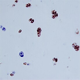
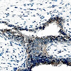
- Immunohistochemical analysis of CHI3L1 in immersion fixed paraffin-embedded sections of human ovarian cancer. Samples were incubated in CHI3L1 polyclonal antibody (Product # PA5-46996) using a dilution of 15 µg/mL overnight at 4 °C. Before incubation with the primary antibody, tissue was subjected to heat-induced epitope retrieval using Antigen Retrieval Reagent-basic. Tissue was stained using the Anti-Goat HRP-DAB Cell & Tissue Staining Kit (brown) and counterstained with hematoxylin (blue). Specific staining was localized to epithelial cells.

- Submitted by
- Invitrogen Antibodies (provider)
- Main image

- Experimental details
- Immunohistochemical analysis of CHI3L1 in immersion fixed paraffin-embedded sections of human cartilage. Samples were incubated in CHI3L1 polyclonal antibody (Product # PA5-46996) using a dilution of 15 µg/mL overnight at 4 °C. Tissue was stained using the Anti-Goat HRP-DAB Cell & Tissue Staining Kit (brown) and counterstained with hematoxylin (blue). Specific staining was localized to chondrocytes.