Explore
 Validate
 Learn
Learn Western blot
Antibody data
- Antibody Data
- Antigen structure
- References [0]
- Comments [0]
- Validations
- Western blot [1]
- Immunocytochemistry [1]
- Immunohistochemistry [1]
- Flow cytometry [1]
Submit
Validation data
Reference
Comment
Report error
- Product number
- NBP2-30069 - Provider product page

- Provider
- Novus Biologicals
- Product name
- Rabbit Polyclonal ARPC1A Antibody
- Antibody type
- Polyclonal
- Description
- Ammonium sulfate precipitation.
- Reactivity
- Human
- Host
- Rabbit
- Isotype
- IgG
- Vial size
- 0.4 ml
- Concentration
- 3.97 mg/ml
- Storage
- Store at 4C short term. Aliquot and store at -20C long term. Avoid freeze-thaw cycles.
No comments: Submit comment
Supportive validation
- Submitted by
- Novus Biologicals (provider)
- Main image
- Experimental details
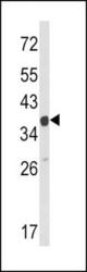
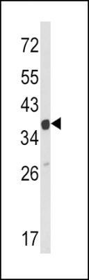
- Western Blot: ARPC1A Antibody [NBP2-30069] - (C-term) (NBP2-30069) in Hela cell line lysates (35ug/lane). ARPC1A (arrow) was detected using the purified Pab.
Supportive validation
- Submitted by
- Novus Biologicals (provider)
- Main image
- Experimental details
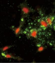
- Immunocytochemistry/Immunofluorescence: ARPC1A Antibody [NBP2-30069] - Immunofluorescence analysis of (C-term) with hela cells. 0.025 mg/ml primary antibody was followed by FITC-conjugated goat anti-rabbit lgG (whole molecule). FITC emits green fluorescence.Red counterstaining is PI.

Supportive validation
- Submitted by
- Novus Biologicals (provider)
- Main image

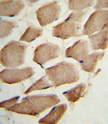
- Experimental details
- Immunohistochemistry-Paraffin: ARPC1A Antibody [NBP2-30069] - Formalin-fixed and paraffin-embedded human skeletal muscle reacted with (C-term), which was peroxidase-conjugated to the secondary antibody, followed by DAB staining. This data demonstrates the use of this antibody for immunohistochemistry; clinical relevance has not been evaluated.
Supportive validation
- Submitted by
- Novus Biologicals (provider)
- Main image
- Experimental details
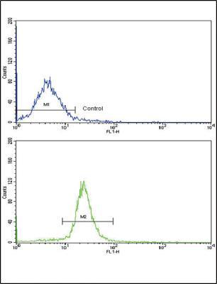
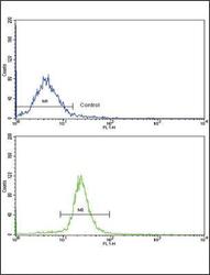
- Flow Cytometry: ARPC1A Antibody [NBP2-30069] - Flow cytometric analysis of WiDr cells using (C-term)(bottom histogram) compared to a negative control cell (top histogram). FITC-conjugated goat-anti-rabbit secondary antibodies were used for the analysis.