 Explore
 Validate
 Learn
Learn Immunocytochemistry
Antibody data
- Antibody Data
- Antigen structure
- References [23]
- Comments [0]
- Validations
- Immunocytochemistry [3]
- Immunohistochemistry [2]
- Flow cytometry [2]
Submit
Validation data
Reference
Comment
Report error
- Product number
- MAB1419 - Provider product page

- Provider
- R&D Systems
- Product name
- Human/Rat Osteocalcin Antibody
- Antibody type
- Monoclonal
- Description
- Protein A or G purified from hybridoma culture supernatant. Detects human Osteocalcin in direct ELISAs.
- Reactivity
- Human, Rat
- Host
- Mouse
- Conjugate
- Unconjugated
- Antigen sequence
P02818- Isotype
- IgG
- Antibody clone number
- 190125
- Vial size
- 100 ug
- Concentration
- LYOPH
- Storage
- Use a manual defrost freezer and avoid repeated freeze-thaw cycles. 12 months from date of receipt, -20 to -70 °C as supplied. 1 month, 2 to 8 °C under sterile conditions after reconstitution. 6 months, -20 to -70 °C under sterile conditions after reconstitution.
Submitted references Comparison of Immunosuppressive and Angiogenic Properties of Human Amnion-Derived Mesenchymal Stem Cells between 2D and 3D Culture Systems.
Osteoblasts are "educated" by crosstalk with metastatic breast cancer cells in the bone tumor microenvironment.
Differential expression patterns of Toll Like Receptors and Interleukin-37 between calcific aortic and mitral valve cusps in humans.
Influences of donor and host age on human muscle-derived stem cell-mediated bone regeneration.
Pharmacological activation of TAZ enhances osteogenic differentiation and bone formation of adipose-derived stem cells.
A biomaterials approach to influence stem cell fate in injectable cell-based therapies.
Rapid Rapamycin-Only Induced Osteogenic Differentiation of Blood-Derived Stem Cells and Their Adhesion to Natural and Artificial Scaffolds.
Collagen type XV and the 'osteogenic status'.
Identification of Multipotent Stem Cells in Human Brain Tissue Following Stroke.
25-Hydroxyvitamin D3 induces osteogenic differentiation of human mesenchymal stem cells.
Transcriptome sequencing wide functional analysis of human mesenchymal stem cells in response to TLR4 ligand.
Immobilized WNT Proteins Act as a Stem Cell Niche for Tissue Engineering.
Ultra-Porous Nanoparticle Networks: A Biomimetic Coating Morphology for Enhanced Cellular Response and Infiltration.
PDL regeneration via cell homing in delayed replantation of avulsed teeth.
Primary osteoblast-like cells from patients with end-stage kidney disease reflect gene expression, proliferation, and mineralization characteristics ex vivo.
Identification of a cell-of-origin for fibroblasts comprising the fibrotic reticulum in idiopathic pulmonary fibrosis.
Bone matrix, cellularity, and structural changes in a rat model with high-turnover osteoporosis induced by combined ovariectomy and a multiple-deficient diet.
Derivation and expansion using only small molecules of human neural progenitors for neurodegenerative disease modeling.
Dilatational band formation in bone.
Age-related changes in rat bone-marrow mesenchymal stem cell plasticity.
The guidance of human mesenchymal stem cell differentiation in vitro by controlled modifications to the cell substrate.
A hybrid coating of biomimetic apatite and osteocalcin.
A hybrid coating of biomimetic apatite and osteocalcin.
Miceli V, Pampalone M, Vella S, Carreca AP, Amico G, Conaldi PG
Stem cells international 2019;2019:7486279
Stem cells international 2019;2019:7486279
Osteoblasts are "educated" by crosstalk with metastatic breast cancer cells in the bone tumor microenvironment.
Kolb AD, Shupp AB, Mukhopadhyay D, Marini FC, Bussard KM
Breast cancer research : BCR 2019 Feb 27;21(1):31
Breast cancer research : BCR 2019 Feb 27;21(1):31
Differential expression patterns of Toll Like Receptors and Interleukin-37 between calcific aortic and mitral valve cusps in humans.
Kapelouzou A, Kontogiannis C, Tsilimigras DI, Georgiopoulos G, Kaklamanis L, Tsourelis L, Cokkinos DV
Cytokine 2019 Apr;116:150-160
Cytokine 2019 Apr;116:150-160
Influences of donor and host age on human muscle-derived stem cell-mediated bone regeneration.
Gao X, Lu A, Tang Y, Schneppendahl J, Liebowitz AB, Scibetta AC, Morris ER, Cheng H, Huard C, Amra S, Wang B, Hall MA, Lowe WR, Huard J
Stem cell research & therapy 2018 Nov 21;9(1):316
Stem cell research & therapy 2018 Nov 21;9(1):316
Pharmacological activation of TAZ enhances osteogenic differentiation and bone formation of adipose-derived stem cells.
Zhu Y, Wu Y, Cheng J, Wang Q, Li Z, Wang Y, Wang D, Wang H, Zhang W, Ye J, Jiang H, Wang L
Stem cell research & therapy 2018 Mar 7;9(1):53
Stem cell research & therapy 2018 Mar 7;9(1):53
A biomaterials approach to influence stem cell fate in injectable cell-based therapies.
Amer MH, Rose FRAJ, Shakesheff KM, White LJ
Stem cell research & therapy 2018 Feb 21;9(1):39
Stem cell research & therapy 2018 Feb 21;9(1):39
Rapid Rapamycin-Only Induced Osteogenic Differentiation of Blood-Derived Stem Cells and Their Adhesion to Natural and Artificial Scaffolds.
Arianna C, Eliana C, Flavio A, Marco R, Giacomo D, Manuel S, Elena B, Alessandra G
Stem cells international 2017;2017:2976541
Stem cells international 2017;2017:2976541
Collagen type XV and the 'osteogenic status'.
Lisignoli G, Lambertini E, Manferdini C, Gabusi E, Penolazzi L, Paolella F, Angelozzi M, Casagranda V, Piva R
Journal of cellular and molecular medicine 2017 Sep;21(9):2236-2244
Journal of cellular and molecular medicine 2017 Sep;21(9):2236-2244
Identification of Multipotent Stem Cells in Human Brain Tissue Following Stroke.
Tatebayashi K, Tanaka Y, Nakano-Doi A, Sakuma R, Kamachi S, Shirakawa M, Uchida K, Kageyama H, Takagi T, Yoshimura S, Matsuyama T, Nakagomi T
Stem cells and development 2017 Jun 1;26(11):787-797
Stem cells and development 2017 Jun 1;26(11):787-797
25-Hydroxyvitamin D3 induces osteogenic differentiation of human mesenchymal stem cells.
Lou YR, Toh TC, Tee YH, Yu H
Scientific reports 2017 Feb 17;7:42816
Scientific reports 2017 Feb 17;7:42816
Transcriptome sequencing wide functional analysis of human mesenchymal stem cells in response to TLR4 ligand.
Kim SH, Das A, Chai JC, Binas B, Choi MR, Park KS, Lee YS, Jung KH, Chai YG
Scientific reports 2016 Jul 22;6:30311
Scientific reports 2016 Jul 22;6:30311
Immobilized WNT Proteins Act as a Stem Cell Niche for Tissue Engineering.
Lowndes M, Rotherham M, Price JC, El Haj AJ, Habib SJ
Stem cell reports 2016 Jul 12;7(1):126-37
Stem cell reports 2016 Jul 12;7(1):126-37
Ultra-Porous Nanoparticle Networks: A Biomimetic Coating Morphology for Enhanced Cellular Response and Infiltration.
Nasiri N, Ceramidas A, Mukherjee S, Panneerselvan A, Nisbet DR, Tricoli A
Scientific reports 2016 Apr 14;6:24305
Scientific reports 2016 Apr 14;6:24305
PDL regeneration via cell homing in delayed replantation of avulsed teeth.
Zhu W, Zhang Q, Zhang Y, Cen L, Wang J
Journal of translational medicine 2015 Nov 14;13:357
Journal of translational medicine 2015 Nov 14;13:357
Primary osteoblast-like cells from patients with end-stage kidney disease reflect gene expression, proliferation, and mineralization characteristics ex vivo.
Pereira RC, Delany AM, Khouzam NM, Bowen RE, Freymiller EG, Salusky IB, Wesseling-Perry K
Kidney international 2015 Mar;87(3):593-601
Kidney international 2015 Mar;87(3):593-601
Identification of a cell-of-origin for fibroblasts comprising the fibrotic reticulum in idiopathic pulmonary fibrosis.
Xia H, Bodempudi V, Benyumov A, Hergert P, Tank D, Herrera J, Braziunas J, Larsson O, Parker M, Rossi D, Smith K, Peterson M, Limper A, Jessurun J, Connett J, Ingbar D, Phan S, Bitterman PB, Henke CA
The American journal of pathology 2014 May;184(5):1369-83
The American journal of pathology 2014 May;184(5):1369-83
Bone matrix, cellularity, and structural changes in a rat model with high-turnover osteoporosis induced by combined ovariectomy and a multiple-deficient diet.
Govindarajan P, Böcker W, El Khassawna T, Kampschulte M, Schlewitz G, Huerter B, Sommer U, Dürselen L, Ignatius A, Bauer N, Szalay G, Wenisch S, Lips KS, Schnettler R, Langheinrich A, Heiss C
The American journal of pathology 2014 Mar;184(3):765-77
The American journal of pathology 2014 Mar;184(3):765-77
Derivation and expansion using only small molecules of human neural progenitors for neurodegenerative disease modeling.
Reinhardt P, Glatza M, Hemmer K, Tsytsyura Y, Thiel CS, Höing S, Moritz S, Parga JA, Wagner L, Bruder JM, Wu G, Schmid B, Röpke A, Klingauf J, Schwamborn JC, Gasser T, Schöler HR, Sterneckert J
PloS one 2013;8(3):e59252
PloS one 2013;8(3):e59252
Dilatational band formation in bone.
Poundarik AA, Diab T, Sroga GE, Ural A, Boskey AL, Gundberg CM, Vashishth D
Proceedings of the National Academy of Sciences of the United States of America 2012 Nov 20;109(47):19178-83
Proceedings of the National Academy of Sciences of the United States of America 2012 Nov 20;109(47):19178-83
Age-related changes in rat bone-marrow mesenchymal stem cell plasticity.
Asumda FZ, Chase PB
BMC cell biology 2011 Oct 12;12:44
BMC cell biology 2011 Oct 12;12:44
The guidance of human mesenchymal stem cell differentiation in vitro by controlled modifications to the cell substrate.
Curran JM, Chen R, Hunt JA
Biomaterials 2006 Sep;27(27):4783-93
Biomaterials 2006 Sep;27(27):4783-93
A hybrid coating of biomimetic apatite and osteocalcin.
Krout A, Wen HB, Hippensteel E, Li P
Journal of biomedical materials research. Part A 2005 Jun 15;73(4):377-87
Journal of biomedical materials research. Part A 2005 Jun 15;73(4):377-87
A hybrid coating of biomimetic apatite and osteocalcin.
Krout A, Wen HB, Hippensteel E, Li P
Journal of biomedical materials research. Part A 2005 Jun 15;73(4):377-87
Journal of biomedical materials research. Part A 2005 Jun 15;73(4):377-87
No comments: Submit comment
Supportive validation
- Submitted by
- R&D Systems (provider)
- Main image

- Experimental details
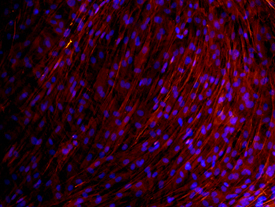
- Osteocalcin in Rat Osteocytes. Osteocalcin was detected in immersion fixed rat osteocytes differentiated from mesenchymal stem cells using Mouse Anti-Human/Rat Osteocalcin Monoclonal Antibody (Catalog # MAB1419) at 10 µg/mL for 3 hours at room temperature. Cells were stained using the NorthernLights™ 557-conjugated Anti-Mouse IgG Secondary Antibody (red; Catalog # NL007) and counterstained with DAPI (blue). View our protocol for Fluorescent ICC Staining of Cells on Coverslips.
- Submitted by
- R&D Systems (provider)
- Main image

- Experimental details
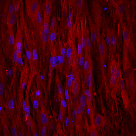
- Osteocalcin in Human Osteocytes. Osteocalcin was detected in human mesenchymal stem cells differentiated into osteocytes using Mouse Anti-Human/Rat Osteocalcin Monoclonal Antibody (Catalog # MAB1419) at 10 µg/mL for 3 hours at room temperature. Cells were stained using the NorthernLights™ 557-conjugated Anti-Mouse IgG Secondary Antibody (red; Catalog # NL007) and counterstained with DAPI (blue). View our protocol for Fluorescent ICC Staining of Cells on Coverslips.
- Submitted by
- R&D Systems (provider)
- Main image
- Experimental details
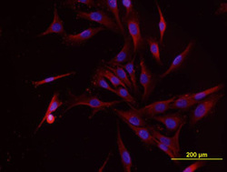
- Osteocalcin in MG-63 Human Cell Line. Osteocalcin was detected in immersion fixed MG-63 human osteosarcoma cell line using Mouse Anti-Human/Rat Osteocalcin Monoclonal Antibody (Catalog # MAB1419) at 10 µg/mL for 3 hours at room temperature. Cells were stained using the NorthernLights™ 557-conjugated Anti-Mouse IgG Secondary Antibody (red; Catalog # NL007) and counterstained with DAPI (blue). View our protocol for Fluorescent ICC Staining of Cells on Coverslips.

Supportive validation
- Submitted by
- R&D Systems (provider)
- Main image

- Experimental details
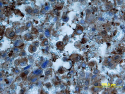
- Osteocalcin in Human Cartilage. Osteocalcin was detected in immersion fixed paraffin-embedded sections of human cartilage using Mouse Anti-Human/Rat Osteocalcin Monoclonal Antibody (Catalog # MAB1419) at 8 µg/mL overnight at 4 °C. Tissue was stained using the Anti-Mouse HRP-DAB Cell & Tissue Staining Kit (brown; Catalog # CTS002) and counterstained with hematoxylin (blue). Specific labeling was localized to the cytoplasm of chondrocytes. View our protocol for Chromogenic IHC Staining of Paraffin-embedded Tissue Sections.

- Submitted by
- R&D Systems (provider)
- Main image
- Experimental details
- Osteocalcin in Human Osteosarcoma. Osteocalcin was detected in immersion fixed paraffin-embedded sections of human osteosarcoma using Mouse Anti-Human/Rat Osteocalcin Monoclonal Antibody (Catalog # MAB1419) at 25 µg/mL overnight at 4 °C. Tissue was stained using the Anti-Mouse HRP-DAB Cell & Tissue Staining Kit (brown; Catalog # CTS002) and counterstained with hematoxylin (blue). View our protocol for Chromogenic IHC Staining of Paraffin-embedded Tissue Sections.

Supportive validation
- Submitted by
- R&D Systems (provider)
- Main image
- Experimental details
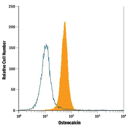
- Detection of Osteocalcin in Saos-2 Human Cell Line by Flow Cytometry. Saos-2 human osteosarcoma cell line was stained with Mouse Anti-Human/Rat Osteocalcin Monoclonal Antibody (Catalog # MAB1419, filled histogram) or isotype control antibody (Catalog # MAB002, open histogram), followed by Allophycocyanin-conjugated Anti-Mouse IgG Secondary Antibody (Catalog # F0101B). To facilitate intracellular staining, cells were fixed with paraformaldehyde and permeabilized with saponin.

- Submitted by
- R&D Systems (provider)
- Main image

- Experimental details
- Detection of Osteocalcin in Human Osteoblasts by Flow Cytometry. Human osteoblasts were stained with Mouse Anti-Human/Rat Osteocalcin Monoclonal Antibody (Catalog # MAB1419, filled histogram) or isotype control antibody (Catalog # MAB002, open histogram), followed by Allophycocyanin-conjugated Anti-Mouse IgG Secondary Antibody (Catalog # F0101B). To facilitate intracellular staining, cells were fixed with paraformaldehyde and permeabilized with saponin.
