Explore
 Validate
 Learn
Learn Western blot
Antibody data
- Antibody Data
- Antigen structure
- References [0]
- Comments [0]
- Validations
- Western blot [4]
- Immunocytochemistry [2]
- Immunohistochemistry [1]
- Flow cytometry [1]
Submit
Validation data
Reference
Comment
Report error
- Product number
- TA302025 - Provider product page

- Provider
- OriGene
- Product name
- Mouse Monoclonal Antibody against SOX2
- Antibody type
- Monoclonal
- Description
- Mouse Monoclonal Antibody against SOX2
- Host
- Mouse
- Conjugate
- Unconjugated
- Epitope
- SOX2
- Isotype
- IgG
- Antibody clone number
- NULL
- Vial size
- 100 µg
- Concentration
- NULL
No comments: Submit comment
Supportive validation
- Submitted by
- OriGene (provider)
- Main image
- Experimental details
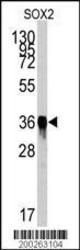
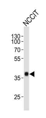
- Western blot analysis of SOX2 Antibody (Cat.#TA302025) by SOX2 recombinant protein. SOX2(arrow) was detected using the purified Mab.
- Validation comment
- WB

- Submitted by
- OriGene (provider)
- Main image
- Experimental details
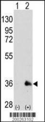
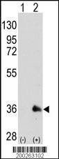
- Western blot analysis of SOX2 (arrow) using mouse monoclonal SOX2 antibody (Cat.#TA302025). 293 cell lysates (2 ug/lane) either nontransfected (Lane 1) or transiently transfected with the SOX2 gene (Lane 2) (Origene Technologies)
- Validation comment
- WB
- Submitted by
- OriGene (provider)
- Main image

- Experimental details
- Western blot analysis of SOX2 Antibody (Cat.#TA302025) by SOX2 recombinant protein. SOX2(arrow) was detected using the purified Mab.
- Validation comment
- WB
- Submitted by
- OriGene (provider)
- Main image

- Experimental details
- Western blot analysis of SOX2 (arrow) using mouse monoclonal SOX2 antibody (Cat.#TA302025). 293 cell lysates (2 ug/lane) either nontransfected (Lane 1) or transiently transfected with the SOX2 gene (Lane 2) (Origene Technologies)
- Validation comment
- WB

Supportive validation
- Submitted by
- OriGene (provider)
- Main image
- Experimental details
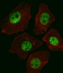
- IF image of A549 cell stained with SOX2 Antibody(Cat#TA302025/SG110310AA).A549 cells were incubated with SOX2 primary antibody (1:25, 1 h at 37?). For secondary antibody, Alexa Fluor? 488 conjugated donkey anti-mouse antibody (green) was used (1:400).Cytoplasmic actin was counterstained with Alexa Fluor? 555 (red) conjugated Phalloidin (7 units/ml).SOX2 immunoreactivity is localized to Nucleus significantly.
- Validation comment
- IF

- Submitted by
- OriGene (provider)
- Main image

- Experimental details
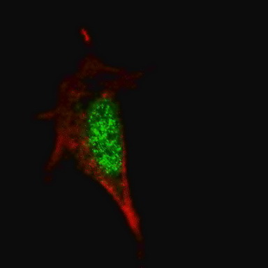
- Fluorescent confocal image of SY5Y cells stained with SOX2 antibody. SY5Y cells were fixed with 4% PFA (20 min), permeabilized with Triton X-100 (0.2%, 30 min). Cells were then incubated with TA302025 SOX2 primary antibody (1:100, 2 h at room temperature). For secondary antibody, Alexa Fluor?? 488 conjugated donkey anti-mouse antibody (green) was used (1:1000, 1h). Note the highly specific localization of the SOX2 mainly to the nucleus.
- Validation comment
- IF
Supportive validation
- Submitted by
- OriGene (provider)
- Main image

- Experimental details
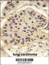
- Formalin-fixed and paraffin-embedded human lung carcinoma tissue reacted with SOX2 Antibody (Cat.#TA302025), which was peroxidase-conjugated to the secondary antibody, followed by DAB staining. This data demonstrates the use of this antibody for immunohistochemistry; clinical relevance has not been evaluated.
- Validation comment
- IHC
Supportive validation
- Submitted by
- OriGene (provider)
- Main image

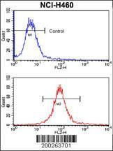
- Experimental details
- Flow cytometric analysis of NCI-H460 cells using SOX2 Monoclonal Antibody (bottom histogram) compared to a negative control cell (top histogram). PE-conjugated goat-anti-mouse secondary antibodies were used for the analysis.
- Validation comment
- FC