Explore
 Validate
 Learn
Learn Immunocytochemistry
Antibody data
- Antibody Data
- Antigen structure
- References [0]
- Comments [0]
- Validations
- Immunocytochemistry [1]
- Flow cytometry [1]
Submit
Validation data
Reference
Comment
Report error
- Product number
- MAB5609-100 - Provider product page

- Provider
- R&D Systems
- Product name
- Human CA125/MUC16 Antibody
- Antibody type
- Monoclonal
- Description
- Protein A or G purified from ascites. Detects human CA125/MUC-16 in direct ELISAs.
- Reactivity
- Human
- Host
- Mouse
- Conjugate
- Unconjugated
- Antigen sequence
Q8WX17- Isotype
- IgG
- Antibody clone number
- 986808
- Vial size
- 100 ug
- Storage
- Use a manual defrost freezer and avoid repeated freeze-thaw cycles. 12 months from date of receipt, -20 to -70 °C as supplied. 1 month, 2 to 8 °C under sterile conditions after reconstitution. 6 months, -20 to -70 °C under sterile conditions after reconstitution.
No comments: Submit comment
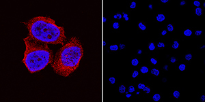
Supportive validation
- Submitted by
- R&D Systems (provider)
- Main image

- Experimental details
- CA125/MUC16 in Hela and U937 Human Cell Lines. CA125/MUC16 was detected in immersion fixed HeLa human cervical epithelial carcinoma cell line (positive control, left panel) and U937 human histiocytic lymphoma cell line (negative control, right panel) using Mouse Anti-Human CA125/MUC16 Monoclonal Antibody (Catalog # MAB5609) at 8 µg/mL for 3 hours at room temperature. Cells were stained using the NorthernLights™ 557-conjugated Anti-Mouse IgG Secondary Antibody (red; Catalog # NL007) and counterstained with DAPI (blue). Specific staining was localized to cytoplasm in HeLa cells. View our protocol for Fluorescent ICC Staining of Cells on Coverslips.
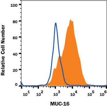
Supportive validation
- Submitted by
- R&D Systems (provider)
- Main image
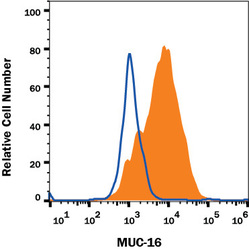
- Experimental details
- Detection of CA125/MUC16 in HeLa Human Cell Line by Flow Cytometry. HeLa human cervical cancer cell line was stained with Mouse Anti-Human CA125/MUC16 Monoclonal Antibody (Catalog # MAB5609, filled histogram) or isotype control antibody (Catalog # MAB003, open histogram), followed by Allophycocyanin-conjugated Anti-Mouse IgG Secondary Antibody (Catalog # F0101B).View our protocol for Staining Membrane-associated Proteins.