Explore
 Validate
 Learn
Learn Western blot
Antibody data
- Antibody Data
- Antigen structure
- References [5]
- Comments [0]
- Validations
- Western blot [1]
- Immunocytochemistry [2]
- Immunohistochemistry [1]
- Flow cytometry [2]
Submit
Validation data
Reference
Comment
Report error
- Product number
- MA5-12954 - Provider product page

- Provider
- Invitrogen Antibodies
- Product name
- CD99 Monoclonal Antibody (HO36-1.1)
- Antibody type
- Monoclonal
- Antigen
- Purifed from natural sources
- Description
- MA5-12954 targets CD99 in WB, IF, FACS and IHC (P) applications and shows reactivity with Human and Rat samples. This antibody is not suitable for Mouse spleen cells in Western blot analysis.
- Antibody clone number
- HO36-1.1
- Concentration
- 0.4 mg/mL
Submitted references Primary sclerosing epithelioid fibrosarcoma of kidney with variant histomorphologic features: report of 2 cases and review of the literature.
Pericardial synovial sarcoma, a potential for misdiagnosis: clinicopathologic and molecular cytogenetic analysis of three cases with literature review.
Extraventricular neurocytomas: a morphological and histogenetic consideration. A study of six cases.
Primary sellar neuroblastoma presenting with syndrome of inappropriate secretion of anti-diuretic hormone.
Uterine carcinosarcoma metastatic to the lung as large-cell neuroendocrine carcinoma with synchronous sarcoid granulomatosis.
Ertoy Baydar D, Kosemehmetoglu K, Aydin O, Bridge JA, Buyukeren B, Aki FT
Diagnostic pathology 2015 Oct 9;10:186
Diagnostic pathology 2015 Oct 9;10:186
Pericardial synovial sarcoma, a potential for misdiagnosis: clinicopathologic and molecular cytogenetic analysis of three cases with literature review.
Cheng Y, Sheng W, Zhou X, Wang J
American journal of clinical pathology 2012 Jan;137(1):142-9
American journal of clinical pathology 2012 Jan;137(1):142-9
Extraventricular neurocytomas: a morphological and histogenetic consideration. A study of six cases.
Agarwal S, Sharma MC, Sarkar C, Suri V, Jain A, Sharma MS, Ailawadhi P, Garg A, Mallick S
Pathology 2011 Jun;43(4):327-34
Pathology 2011 Jun;43(4):327-34
Primary sellar neuroblastoma presenting with syndrome of inappropriate secretion of anti-diuretic hormone.
Radotra B, Apostolopoulos V, Sandison A, Hatfield EC, Mendoza N, Moss J, Mehta A, Glaser M, Meeran K, Roncaroli F
Endocrine pathology 2010 Dec;21(4):266-73
Endocrine pathology 2010 Dec;21(4):266-73
Uterine carcinosarcoma metastatic to the lung as large-cell neuroendocrine carcinoma with synchronous sarcoid granulomatosis.
Froio E, D'Adda T, Fellegara G, Martella E, Caruana P, Pruneri G, Pesci A, Rindi G
Lung cancer (Amsterdam, Netherlands) 2009 Jun;64(3):371-7
Lung cancer (Amsterdam, Netherlands) 2009 Jun;64(3):371-7
No comments: Submit comment
Supportive validation
- Submitted by
- Invitrogen Antibodies (provider)
- Main image

- Experimental details
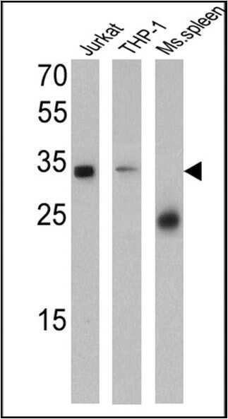
- Western blot analysis of CD99 was performed by loading 25 µg of Jurkat (Lane 1), THP-1 (Lane 2), and Mouse spleen (Lane 3) cell lysates and a molecular weight protein ladder onto an SDS polyacrylamide gel. Proteins were transferred to a PVDF membrane and blocked with a blocking buffer at 4ºC overnight. The membrane was probed with a CD99 monoclonal antibody (Product # MA5-12954) at a dilution of 1:200 (Jurkat and Mouse spleen) and 1:100 (THP-1) overnight at 4°C, washed in TBST, and probed with an HRP-conjugated secondary antibody for 1 hr at room temperature in the dark. Results show a band at 32 kDa in Jurkat and THP-1 cell lysates.
Supportive validation
- Submitted by
- Invitrogen Antibodies (provider)
- Main image
- Experimental details
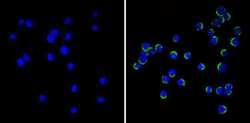
- Immunofluorescent analysis of CD99 (green) showing staining in the membrane of Jurkat cells (right) compared to a negative control without primary antibody (left). Formalin-fixed cells were permeabilized with 0.1% Triton X-100 in TBS for 5-10 minutes and blocked with 3% BSA-PBS for 30 minutes at room temperature. Cells were probed with a CD99 monoclonal antibody (Product # MA5-12954) in 3% BSA-PBS at a dilution of 1:20 and incubated overnight at 4ºC in a humidified chamber. Cells were washed with PBST and incubated with a DyLight-conjugated secondary antibody in PBS at room temperature in the dark. F-actin (red) was stained with a fluorescent red phalloidin and nuclei (blue) were stained with Hoechst or DAPI. Images were taken at a magnification of 60x.

- Submitted by
- Invitrogen Antibodies (provider)
- Main image
- Experimental details
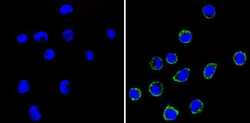
- Immunofluorescent analysis of CD99 (green) showing staining in the membrane of THP-1 cells (right) compared to a negative control without primary antibody (left). Formalin-fixed cells were permeabilized with 0.1% Triton X-100 in TBS for 5-10 minutes and blocked with 3% BSA-PBS for 30 minutes at room temperature. Cells were probed with a CD99 monoclonal antibody (Product # MA5-12954) in 3% BSA-PBS at a dilution of 1:20 and incubated overnight at 4ºC in a humidified chamber. Cells were washed with PBST and incubated with a DyLight-conjugated secondary antibody in PBS at room temperature in the dark. F-actin (red) was stained with a fluorescent red phalloidin and nuclei (blue) were stained with Hoechst or DAPI. Images were taken at a magnification of 60x.

Supportive validation
- Submitted by
- Invitrogen Antibodies (provider)
- Main image

- Experimental details
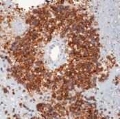
- Formalin-fixed, paraffin-embedded human Ewings sarcoma stained with CD99 antibody using peroxidase-conjugate and DAB chromogen. Note cell membrane staining of tumor cells.
Supportive validation
- Submitted by
- Invitrogen Antibodies (provider)
- Main image
- Experimental details
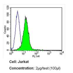
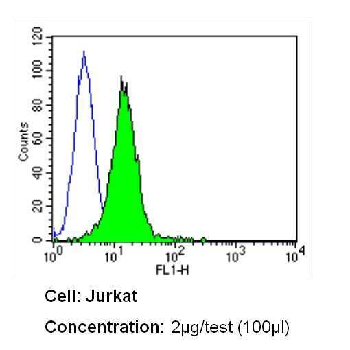
- Flow cytometry analysis of CD99 in Jurkat cells (green) compared to an isotype control (blue). Cells were harvested, adjusted to a concentration of 1-5x10^6 cells/mL, fixed with 2% paraformaldehyde and washed with PBS. Cells were blocked with a 2% solution of BSA-PBS for 30 min at room temperature and incubated with a CD99 monoclonal antibody (Product # MA5-12954) at a dilution of 2 µg/test for 60 min at room temperature. Cells were then incubated for 40 min at room temperature in the dark using a Dylight 488-conjugated secondary antibody and re-suspended in PBS for FACS analysis.
- Submitted by
- Invitrogen Antibodies (provider)
- Main image
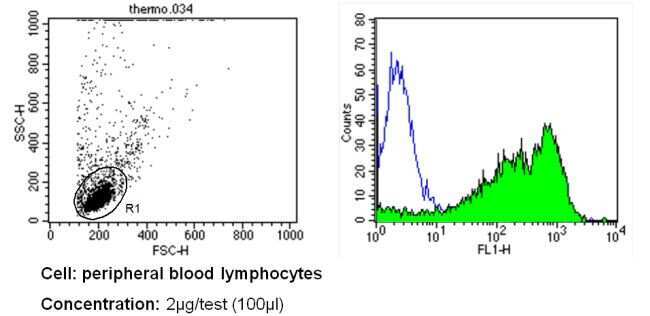
- Experimental details
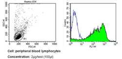
- Flow cytometry analysis of CD99 in PBMC cells (green) compared to an isotype control (blue). Human blood was collected, combined with a hydrophilic polysaccharide, centrifuged, transferred to a conical tube and washed with PBS. 50 µL of cell solution was added to each tube at a dilution of 2x10^7 cells/mL, followed by the addition of 50 µL of isotype control and primary antibody (Product # MA5-12954) at a dilution of 2 µg/test. Cells were incubated for 30 min at 4ºC and washed with a cell buffer, followed by incubation with a DyLight 488-conjugated secondary antibody for 30 min at 4ºC in the dark. FACS analysis was performed using 400 µL of cell buffer.