 Explore
 Validate
 Learn
Learn Western blot
 Immunohistochemistry
Antibody data
- Antibody Data
- Antigen structure
- References [0]
- Comments [0]
- Validations
- Western blot [2]
- Immunohistochemistry [16]
Submit
Validation data
Reference
Comment
Report error
- Product number
- HPA019472 - Provider product page

- Provider
- Atlas Antibodies
- Proper citation
- Atlas Antibodies Cat#HPA019472, RRID:AB_1857360
- Product name
- Anti-SST
- Antibody type
- Polyclonal
- Reactivity
- Human, Mouse
- Host
- Rabbit
- Conjugate
- Unconjugated
- Antigen sequence
APSDPRLRQFLQKSLAAAAGKQELAKYFLAELLSE
PNQTENDALEPEDLSQAAEQDEMRLELQRSANSNP
AMAPRERKAGCKN- Isotype
- IgG
- Vial size
- 100 µl
- Storage
- Store at +4°C for short term storage. Long time storage is recommended at -20°C.
No comments: Submit comment
Supportive validation
- Submitted by
- Atlas Antibodies (provider)
- Main image

- Experimental details
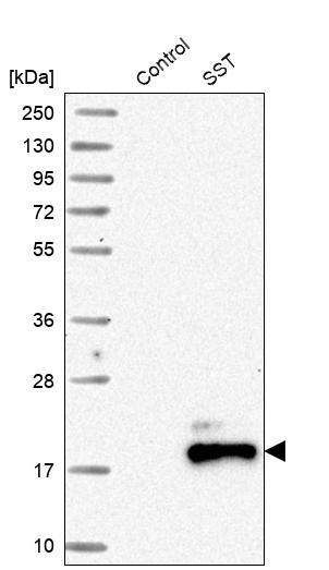
- Western blot analysis in control (vector only transfected HEK293T lysate) and SST over-expression lysate (Co-expressed with a C-terminal myc-DDK tag (~3.1 kDa) in mammalian HEK293T cells, LY420765).
- Sample type
- HUMAN
- Submitted by
- Atlas Antibodies (provider)
- Main image
- Experimental details
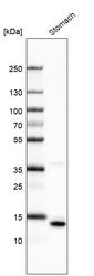
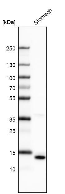
- Western blot analysis in human stomach tissue.
Enhanced validation
Supportive validation
- Submitted by
- Atlas Antibodies (provider)
- Enhanced method
- Orthogonal validation
- Main image

- Experimental details
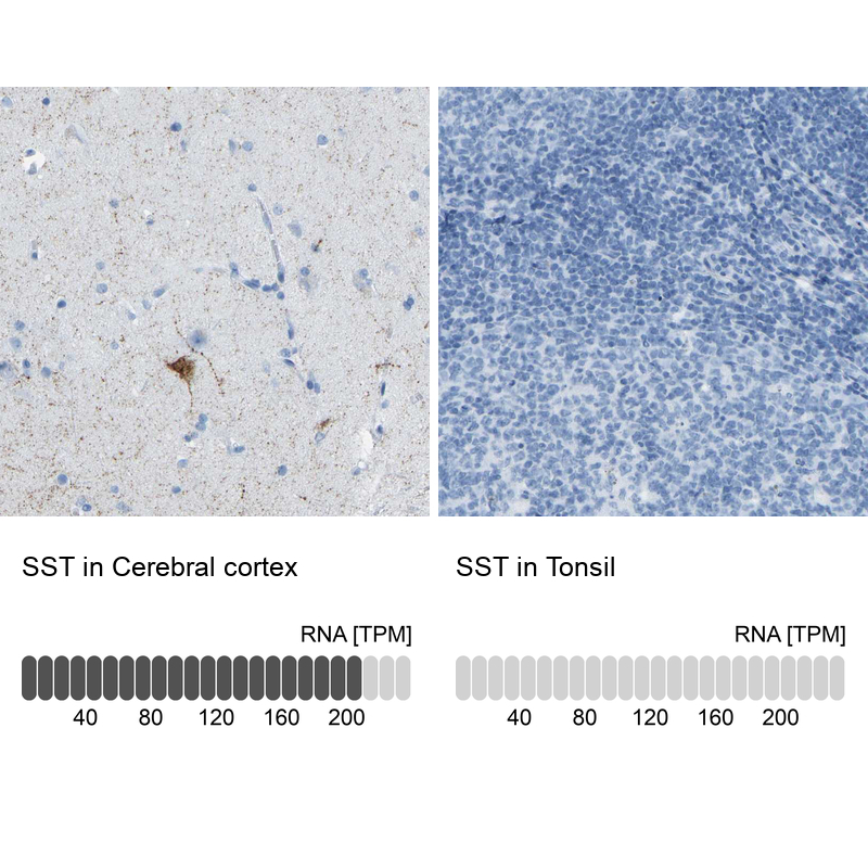
- Immunohistochemistry analysis in human cerebral cortex and tonsil tissues using HPA019472 antibody. Corresponding SST RNA-seq data are presented for the same tissues.
- Sample type
- HUMAN
Supportive validation
- Submitted by
- Atlas Antibodies (provider)
- Main image

- Experimental details
- Immunohistochemical staining of human stomach shows strong positivity in enteroendocrine cells.

- Submitted by
- Atlas Antibodies (provider)
- Main image

- Experimental details
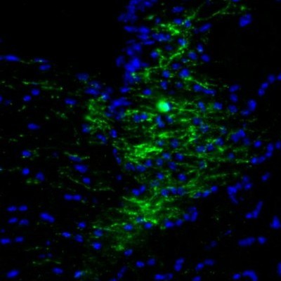
- Immunofluorescence staining of mouse hypothalamus shows immunopositive processes in the central anterior hypothalamic area.
- Submitted by
- Atlas Antibodies (provider)
- Main image
- Experimental details
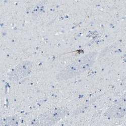
- Immunohistochemical staining of human cerebral cortex shows selective positivity in subsets of neurons.
- Submitted by
- Atlas Antibodies (provider)
- Main image

- Experimental details
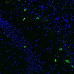
- Immunofluorescence staining of mouse lateral septum shows immunoreactivity in a subset of neurons.

- Submitted by
- Atlas Antibodies (provider)
- Main image
- Experimental details
- Immunofluorescence staining of mouse stria terminalis shows selective positivity in neuronal processes.
- Submitted by
- Atlas Antibodies (provider)
- Main image
- Experimental details
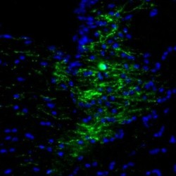
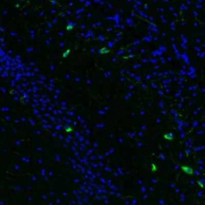
- Immunofluorescence staining of mouse hippocampus shows immunoreactivity in a subset of interneurons in the CA2/ stratum oriens.
- Submitted by
- Atlas Antibodies (provider)
- Main image
- Experimental details
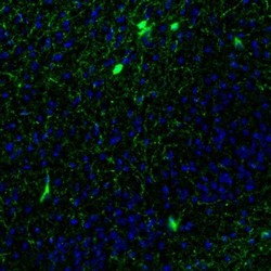
- Immunofluorescence staining of mouse piriform area shows positivity in a subset of interneuron-like cells.

- Submitted by
- Atlas Antibodies (provider)
- Main image
- Experimental details
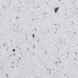
- Immunohistochemical staining of human lateral ventricle wall shows cytoplasmic immunoreactivity in single neurons.
- Submitted by
- Atlas Antibodies (provider)
- Main image
- Experimental details
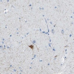
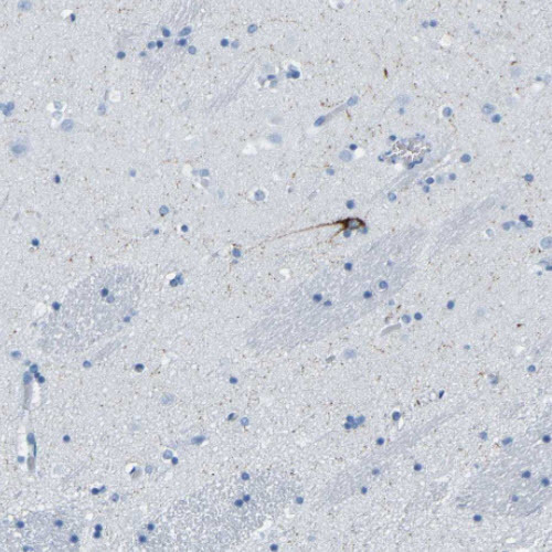
- Immunohistochemical staining of human cerebral cortex shows cytoplasmic positivity in single neurons.
- Submitted by
- Atlas Antibodies (provider)
- Main image

- Experimental details
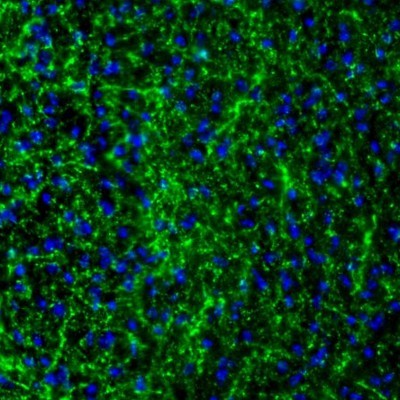
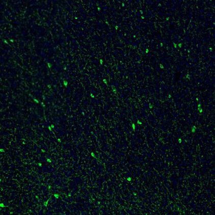
- Immunofluorescence staining of mouse brain shows strong positivity in a subset of neurons in the cerebral cortex.
- Sample type
- MOUSE
- Submitted by
- Atlas Antibodies (provider)
- Main image

- Experimental details
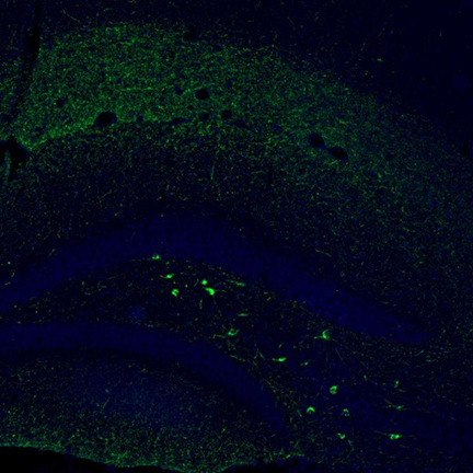
- Immunofluorescence staining of mouse dentate gyrus shows moderate to strong positivity in a subset of neurons in the polymorph layer.
- Sample type
- MOUSE
- Submitted by
- Atlas Antibodies (provider)
- Main image
- Experimental details
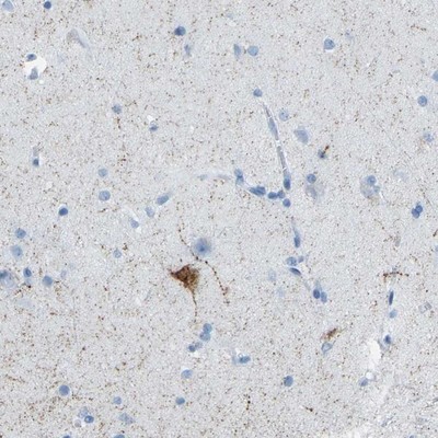
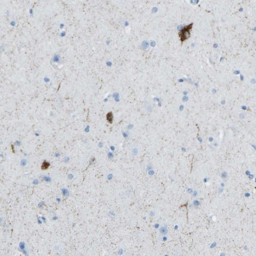
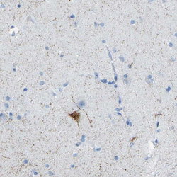
- Immunohistochemical staining of human cerebral cortex shows moderate cytoplasmic positivity in neurons, as well as immunoreactivity in neural processes.
- Sample type
- HUMAN

- Submitted by
- Atlas Antibodies (provider)
- Main image

- Experimental details
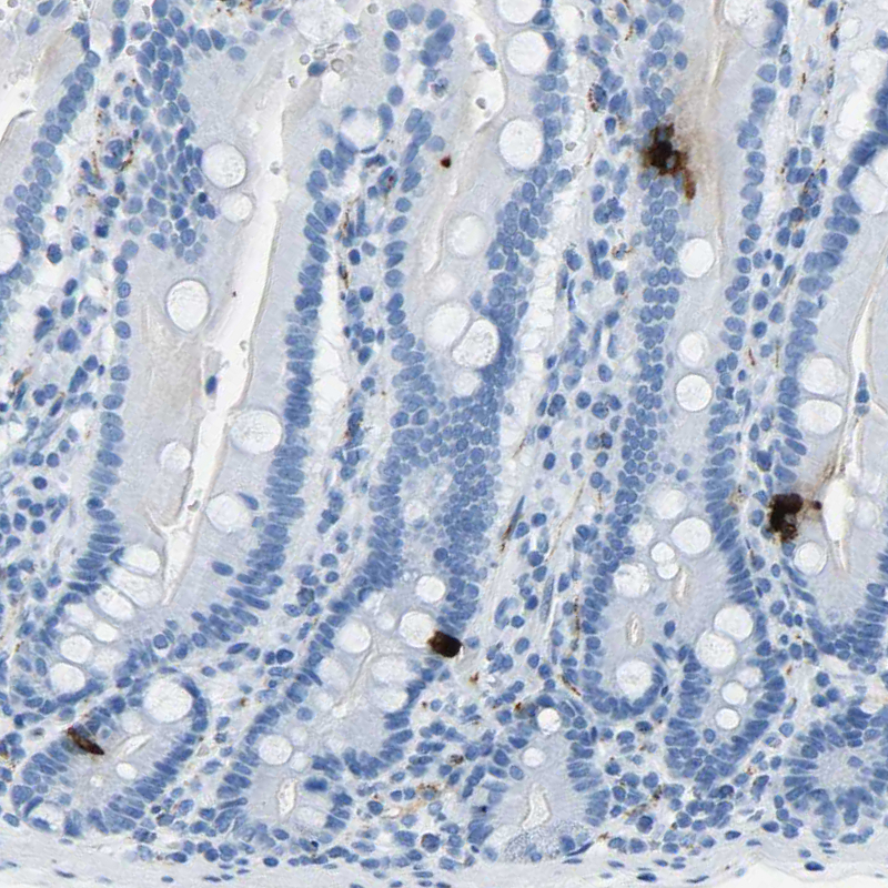
- Immunohistochemical staining of human duodenum shows strong cytoplasmic positivity in neuroendocrine cells.
- Sample type
- HUMAN
- Submitted by
- Atlas Antibodies (provider)
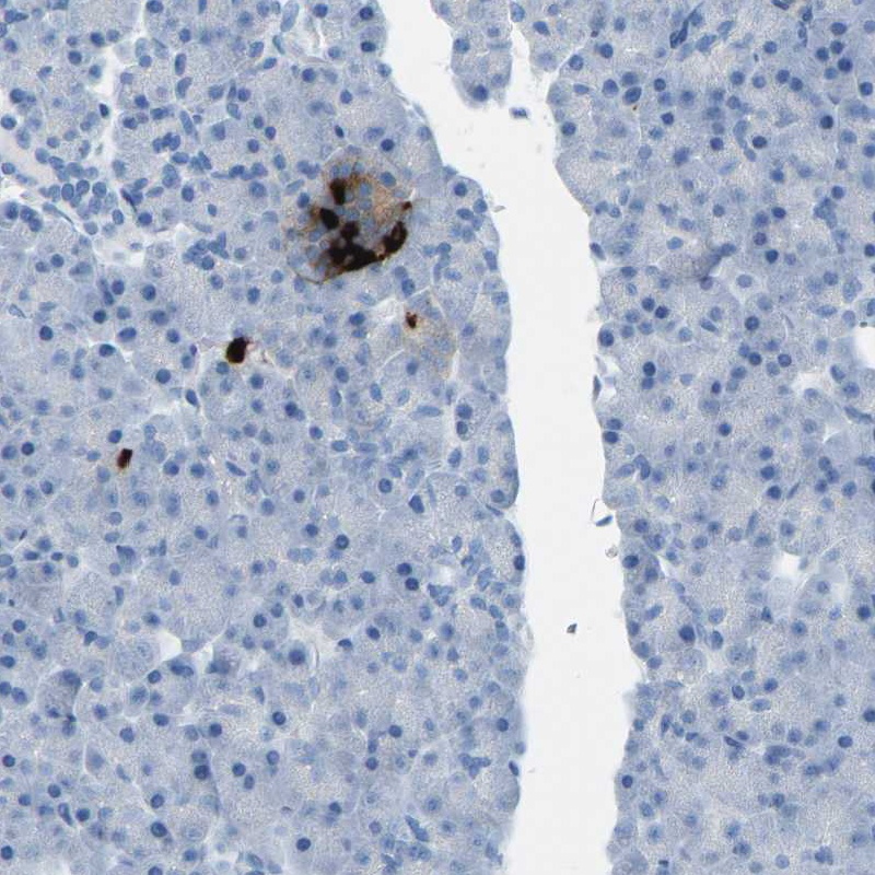
- Main image
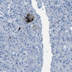
- Experimental details
- Immunohistochemical staining of human pancreas shows strong cytoplasmic positivity in a subset of cells in islets of Langerhans.
- Sample type
- HUMAN
- Submitted by
- Atlas Antibodies (provider)
- Main image
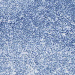
- Experimental details
- Immunohistochemical staining of human tonsil shows no positivity in lymphoid cells as expected.
- Sample type
- HUMAN
