Explore
 Validate
 Learn
LearnNB200-108
antibody from Novus Biologicals
Targeting: MYC
bHLHe39, c-Myc, MYCC
 Western blot
 ELISA Immunoprecipitation Immunohistochemistry Flow cytometry Chromatin Immunoprecipitation
Antibody data
- Antibody Data
- Antigen structure
- References [9]
- Comments [0]
- Validations
- Western blot [1]
- Immunohistochemistry [1]
- Flow cytometry [4]
Submit
Validation data
Reference
Comment
Report error
- Product number
- NB200-108 - Provider product page

- Provider
- Novus Biologicals
- Proper citation
- Novus Cat#NB200-108, RRID:AB_10078595
- Product name
- Mouse Monoclonal c-Myc Antibody
- Antibody type
- Monoclonal
- Description
- Protein A or G purified.
- Reactivity
- Human, Mouse, Rat, Chicken/Avian, Yeast
- Host
- Mouse
- Isotype
- IgG
- Vial size
- 0.1 ml
- Concentration
- 1.0 mg/ml
- Storage
- Store at 4C short term. Aliquot and store at -20C long term. Avoid freeze-thaw cycles.
Submitted references Hormetic dose response to (L)-ascorbic acid as an anti-cancer drug in colorectal cancer cell lines according to SVCT-2 expression.
A nontranscriptional role for HIF-1α as a direct inhibitor of DNA replication.
Nucleolar localization of hepatic c-Myc: a potential mechanism for c-Myc regulation.
Activation of the Kaposi's sarcoma-associated herpesvirus major latency locus by the lytic switch protein RTA (ORF50).
Global position and recruitment of HATs and HDACs in the yeast genome.
The Yaf9 component of the SWR1 and NuA4 complexes is required for proper gene expression, histone H4 acetylation, and Htz1 replacement near telomeres.
Interdependent nuclear accumulation of budding yeast Cdt1 and Mcm2-7 during G1 phase.
Oncogenes and male breast carcinoma: c-erbB-2 and p53 coexpression predicts a poor survival.
Association of the class V myosin Myo4p with a localised messenger RNA in budding yeast depends on She proteins.
Cho S, Chae JS, Shin H, Shin Y, Song H, Kim Y, Yoo BC, Roh K, Cho S, Kil EJ, Byun HS, Cho SH, Park S, Lee S, Yeom CH
Scientific reports 2018 Jul 27;8(1):11372
Scientific reports 2018 Jul 27;8(1):11372
A nontranscriptional role for HIF-1α as a direct inhibitor of DNA replication.
Hubbi ME, Kshitiz, Gilkes DM, Rey S, Wong CC, Luo W, Kim DH, Dang CV, Levchenko A, Semenza GL
Science signaling 2013 Feb 12;6(262):ra10
Science signaling 2013 Feb 12;6(262):ra10
Nucleolar localization of hepatic c-Myc: a potential mechanism for c-Myc regulation.
Sanders JA, Gruppuso PA
Biochimica et biophysica acta 2005 Mar 22;1743(1-2):141-50
Biochimica et biophysica acta 2005 Mar 22;1743(1-2):141-50
Activation of the Kaposi's sarcoma-associated herpesvirus major latency locus by the lytic switch protein RTA (ORF50).
Matsumura S, Fujita Y, Gomez E, Tanese N, Wilson AC
Journal of virology 2005 Jul;79(13):8493-505
Journal of virology 2005 Jul;79(13):8493-505
Global position and recruitment of HATs and HDACs in the yeast genome.
Robert F, Pokholok DK, Hannett NM, Rinaldi NJ, Chandy M, Rolfe A, Workman JL, Gifford DK, Young RA
Molecular cell 2004 Oct 22;16(2):199-209
Molecular cell 2004 Oct 22;16(2):199-209
The Yaf9 component of the SWR1 and NuA4 complexes is required for proper gene expression, histone H4 acetylation, and Htz1 replacement near telomeres.
Zhang H, Richardson DO, Roberts DN, Utley R, Erdjument-Bromage H, Tempst P, Côté J, Cairns BR
Molecular and cellular biology 2004 Nov;24(21):9424-36
Molecular and cellular biology 2004 Nov;24(21):9424-36
Interdependent nuclear accumulation of budding yeast Cdt1 and Mcm2-7 during G1 phase.
Tanaka S, Diffley JF
Nature cell biology 2002 Mar;4(3):198-207
Nature cell biology 2002 Mar;4(3):198-207
Oncogenes and male breast carcinoma: c-erbB-2 and p53 coexpression predicts a poor survival.
Pich A, Margaria E, Chiusa L
Journal of clinical oncology : official journal of the American Society of Clinical Oncology 2000 Aug;18(16):2948-56
Journal of clinical oncology : official journal of the American Society of Clinical Oncology 2000 Aug;18(16):2948-56
Association of the class V myosin Myo4p with a localised messenger RNA in budding yeast depends on She proteins.
Münchow S, Sauter C, Jansen RP
Journal of cell science 1999 May;112 ( Pt 10):1511-8
Journal of cell science 1999 May;112 ( Pt 10):1511-8
No comments: Submit comment
Supportive validation
- Submitted by
- Novus Biologicals (provider)
- Main image
- Experimental details
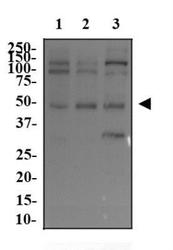
- Western Blot: c-Myc Antibody (9E11) [NB200-108] - Whole cell protein from PC3 (lane 1), U-2 OS (lane 2) and mouse testis (lane 3) was separated on a 12% gel by SDS-PAGE, transferred to PVDF membrane and blocked in 5% non-fat milk in TBST. The membrane was probed with 2.0 ug/ml anti-c-Myc in 1% milk, and detected with an anti-mouse HRP secondary antibody using chemiluminescence.

Supportive validation
- Submitted by
- Novus Biologicals (provider)
- Main image

- Experimental details
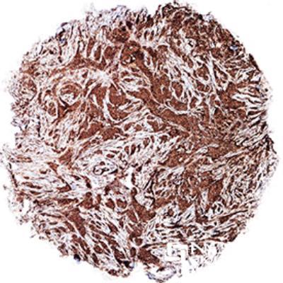
- Immunohistochemistry-Paraffin: c-Myc Antibody (9E11) [NB200-108] - c-Myc was detected in immersion fixed paraffin-embedded sections of human breast cancer using anti-human mouse monoclonal antibody (Catalog # NB200-108, clone 9E11) at 1:600 dilution overnight at 4C. Tissue was stained using the VisuCyte anti-mouse HRP polymer detection reagent (Catalog # VC001) with DAB chromogen (brown) and counterstained with hematoxylin (blue).Images may not be copied, printed or otherwise disseminated without express written permission of Novus Biologicals a bio-techne brand.
Supportive validation
- Submitted by
- Novus Biologicals (provider)
- Main image

- Experimental details
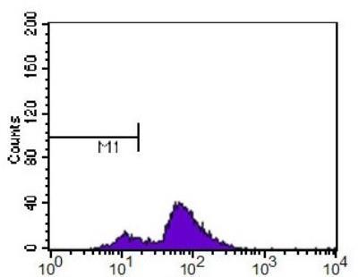
- Flow Cytometry: c-Myc Antibody (9E11) [NB200-108] - c-Myc antibody was tested at 1:400 in HL-60 cells using an Alexa Fluor 488 secondary (shown in purple). M1 is defined by unstained cells.
- Submitted by
- Novus Biologicals (provider)
- Main image
- Experimental details
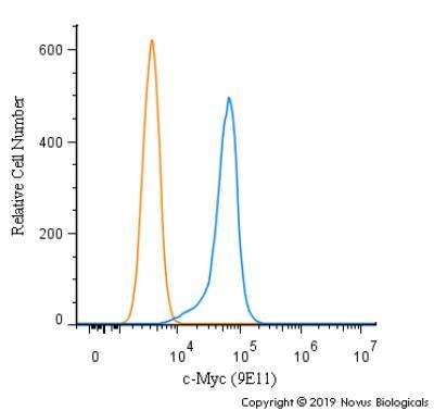
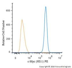
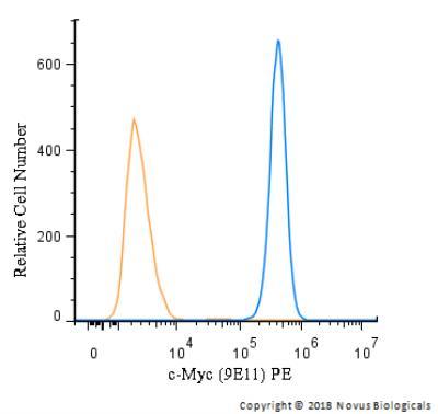
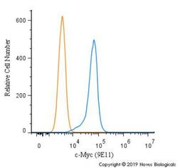
- Flow Cytometry: c-Myc Antibody (9E11) [NB200-108] - An intracellular stain was performed on U-937 cells with c-Myc Antibody (9E11) NB200-108PE (blue) and a matched isotype control (orange). Cells were fixed with 4% PFA and then permeabilized with 0.1% saponin. Cells were incubated in an antibody dilution of 2.5 ug/mL for 30 minutes at room temperature. Both antibodies were conjugated to Phycoerthrin.
- Submitted by
- Novus Biologicals (provider)
- Main image
- Experimental details
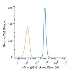
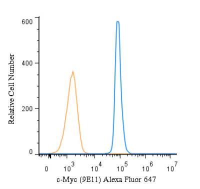
- Flow (Intracellular): c-Myc Antibody (9E11) [NB200-108] - An intracellular stain was performed on U-937 cells with c-Myc Antibody (9E11) NB200-108AF647 (blue) and a matched isotype control (orange). Cells were fixed with 4% PFA and then permeabilized with 0.1% saponin. Cells were incubated in an antibody dilution of 2.5 ug/mL for 30 minutes at room temperature. Both antibodies were conjugated to Alexa Fluor 647.
- Submitted by
- Novus Biologicals (provider)
- Main image
- Experimental details
- Flow Cytometry: c-Myc Antibody (9E11) [NB200-108] - An intracellular stain was performed on U-937 cells with c-Myc Antibody [9E11] NB200-108 (blue) and a matched isotype control (orange). Cells were fixed with 4% PFA and then permeabilized with 0.1% saponin. Cells were incubated in an antibody dilution of 1.0 ug/mL for 30 minutes at room temperature, followed by Mouse IgG (H+L) Cross-Adsorbed Secondary Antibody.