 Explore
 Validate
 Learn
Learn Western blot
 Immunocytochemistry
Antibody data
- Antibody Data
- Antigen structure
- References [6]
- Comments [0]
- Validations
- Western blot [3]
- Immunohistochemistry [2]
Submit
Validation data
Reference
Comment
Report error
- Product number
- NB110-60011 - Provider product page

- Provider
- Novus Biologicals
- Proper citation
- Novus Cat#NB110-60011, RRID:AB_905863
- Product name
- Mouse Monoclonal WT1 Antibody
- Antibody type
- Monoclonal
- Description
- Protein G purified.
- Reactivity
- Human, Mouse
- Host
- Mouse
- Isotype
- IgG
- Vial size
- 0.1 ml
- Concentration
- 1.0 mg/ml
- Storage
- Aliquot and store at -20C or -80C. Avoid freeze-thaw cycles.
Submitted references Podocyte-Specific Loss of Krüppel-Like Factor 6 Increases Mitochondrial Injury in Diabetic Kidney Disease.
Hypoxia-sensitive epigenetic regulation of an antisense-oriented lncRNA controls WT1 expression in myeloid leukemia cells.
BAMBI elimination enhances alternative TGF-β signaling and glomerular dysfunction in diabetic mice.
A preclinical mouse model of glioma with an alternative mechanism of telomere maintenance (ALT).
WT1 protein directly regulates expression of vascular endothelial growth factor and is a mediator of tumor response to hypoxia.
Characterization of monoclonal antibodies directed to the amino-terminus of the WT1, Wilms' tumor suppressor protein.
Horne SJ, Vasquez JM, Guo Y, Ly V, Piret SE, Leonardo AR, Ling J, Revelo MP, Bogenhagen D, Yang VW, He JC, Mallipattu SK
Diabetes 2018 Nov;67(11):2420-2433
Diabetes 2018 Nov;67(11):2420-2433
Hypoxia-sensitive epigenetic regulation of an antisense-oriented lncRNA controls WT1 expression in myeloid leukemia cells.
McCarty G, Loeb DM
PloS one 2015;10(3):e0119837
PloS one 2015;10(3):e0119837
BAMBI elimination enhances alternative TGF-β signaling and glomerular dysfunction in diabetic mice.
Fan Y, Li X, Xiao W, Fu J, Harris RC, Lindenmeyer M, Cohen CD, Guillot N, Baron MH, Wang N, Lee K, He JC, Schlondorff D, Chuang PY
Diabetes 2015 Jun;64(6):2220-33
Diabetes 2015 Jun;64(6):2220-33
A preclinical mouse model of glioma with an alternative mechanism of telomere maintenance (ALT).
Jeitany M, Pineda JR, Liu Q, Porreca RM, Hoffschir F, Desmaze C, Silvestre DC, Mailliet P, Junier MP, Londoño-Vallejo A, Ségal-Bendirdjian E, Chneiweiss H, Boussin FD
International journal of cancer 2015 Apr 1;136(7):1546-58
International journal of cancer 2015 Apr 1;136(7):1546-58
WT1 protein directly regulates expression of vascular endothelial growth factor and is a mediator of tumor response to hypoxia.
McCarty G, Awad O, Loeb DM
The Journal of biological chemistry 2011 Dec 23;286(51):43634-43
The Journal of biological chemistry 2011 Dec 23;286(51):43634-43
Characterization of monoclonal antibodies directed to the amino-terminus of the WT1, Wilms' tumor suppressor protein.
Rauscher FJ 3rd, Morris JF, Fredericks WJ, Lopez-Guisa J, Balakrishnan C, Jost M, Herlyn M, Rodeck U
Hybridoma 1998 Apr;17(2):191-8
Hybridoma 1998 Apr;17(2):191-8
No comments: Submit comment
Supportive validation
- Submitted by
- Novus Biologicals (provider)
- Main image

- Experimental details
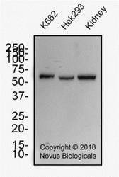
- Simple Western: WT1 Antibody (6F-H2) [NB110-60011] - Lane view shows a specific band for WT1 in 0.5 mg/ml of Hek293 lysate. This experiment was performed under reducing conditions using the 12-230 kDa separation system.

- Submitted by
- Novus Biologicals (provider)
- Main image
- Experimental details
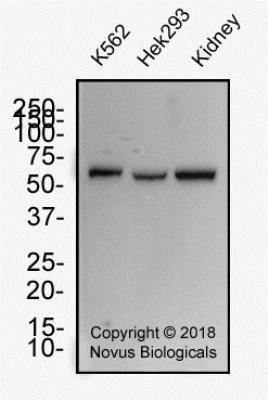
- Western Blot: WT1 Antibody (6F-H2) [NB110-60011] - Whole cell protein from human K562, HEK293 and kidney tissue was separated on a 12% gel by SDS-PAGE, transferred to PVDF membrane and blocked in 5% non-fat milk in TBST. The membrane was probed with 2.0 ug/ml anti-WT1 in block buffer and detected with an anti-mouse HRP secondary antibody using chemiluminescence.
- Submitted by
- Novus Biologicals (provider)
- Main image

- Experimental details
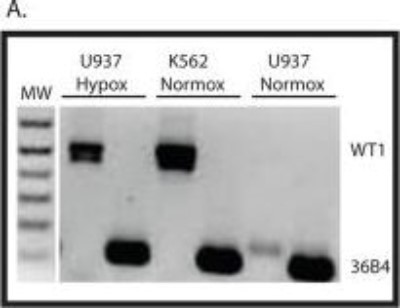
- Western Blot: WT1 Antibody (6F-H2) [NB110-60011] - Hypomethylation of the Intron 1 CpG island results in WT1 expression. RNA was isolated from K562 and U937 cells growing under atmospheric conditions (Normox) and from U937 cells growing in 1% O2 (Hypox) and was analyzed by RT-PCR using primers that span exon 5 of the WT1 mRNA. Ribosomal RNA 36B4 is used as a positive control, and molecular weight markers (MW) are shown. Image collected and cropped by CiteAb from the following publication (http://dx.plos.org/10.1371/journal.pone.0119837), licensed under a CC-BY licence.
Supportive validation
- Submitted by
- Novus Biologicals (provider)
- Main image
- Experimental details
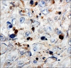
- Immunohistochemistry: WT1 Antibody (6F-H2) [NB110-60011] - Analysis of Wilms Tumor 1 in human renal cancer using DAB with hematoxylin counterstain.

- Submitted by
- Novus Biologicals (provider)
- Main image
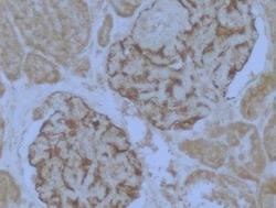
- Experimental details
- Immunohistochemistry-Paraffin: WT1 Antibody (6F-H2) [NB110-60011] - Staining of human kidney glomerulus tissue. Heat mediated antigen retrieval was performed by heating in citrate buffer (pH 6) at 95C for 20 minutes. Image from verified customer review.
