Explore
 Validate
 Learn
Learn Western blot
Antibody data
- Antibody Data
- Antigen structure
- References [3]
- Comments [0]
- Validations
- Western blot [4]
- Immunocytochemistry [1]
- Immunohistochemistry [6]
- Other assay [1]
Submit
Validation data
Reference
Comment
Report error
- Product number
- PA5-85669 - Provider product page

- Provider
- Invitrogen Antibodies
- Product name
- Calbindin D28K Polyclonal Antibody
- Antibody type
- Polyclonal
- Antigen
- Recombinant full-length protein
- Description
- Keep as concentrated solution.
- Concentration
- 1.12 mg/mL
Submitted references Peroxisomal L-bifunctional protein (EHHADH) deficiency causes male-specific kidney hypertrophy and proximal tubular injury in mice.
Cerebellar Kv3.3 potassium channels activate TANK-binding kinase 1 to regulate trafficking of the cell survival protein Hax-1.
Cellular, Molecular and Biochemical Impacts of Silver Nanoparticles on Rat Cerebellar Cortex.
Ranea-Robles P, Portman K, Bender A, Lee K, He JC, Mulholland DJ, Argmann C, Houten SM
Kidney360 2021 Sep;2(9):1441-1454
Kidney360 2021 Sep;2(9):1441-1454
Cerebellar Kv3.3 potassium channels activate TANK-binding kinase 1 to regulate trafficking of the cell survival protein Hax-1.
Zhang Y, Varela L, Szigeti-Buck K, Williams A, Stoiljkovic M, Šestan-Peša M, Henao-Mejia J, D'Acunzo P, Levy E, Flavell RA, Horvath TL, Kaczmarek LK
Nature communications 2021 Mar 19;12(1):1731
Nature communications 2021 Mar 19;12(1):1731
Cellular, Molecular and Biochemical Impacts of Silver Nanoparticles on Rat Cerebellar Cortex.
Mohamed EM, Kattaia AAA, Abdul-Maksoud RS, Abd El-Baset SA
Cells 2020 Dec 22;10(1)
Cells 2020 Dec 22;10(1)
No comments: Submit comment
Supportive validation
- Submitted by
- Invitrogen Antibodies (provider)
- Main image
- Experimental details
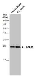
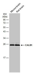
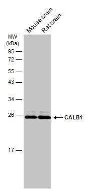
- Western blot analysis of Calbindin D28K in various tissue extracts using Calbindin D28K polyclonal antibody (Product # PA5-85669) using 50 µg of sample at a dilution of 1:1000. Sample was then incubated with HRP-conjugated anti-rabbit IgG secondary antibody. Prior to incubation with primary antibody, the sample was separated on 12% SDS-PAGE.

- Submitted by
- Invitrogen Antibodies (provider)
- Main image
- Experimental details
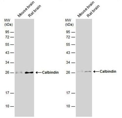
- Western blot analysis of Calbindin D28K in various tissue extracts using Calbindin D28K polyclonal antibody (Product # PA5-85669) using 50 µg of sample at a dilution of 1:1000. Sample was then incubated with HRP-conjugated anti-rabbit IgG secondary antibody. Prior to incubation with primary antibody, the sample was separated on 12% SDS-PAGE.
- Submitted by
- Invitrogen Antibodies (provider)
- Main image
- Experimental details
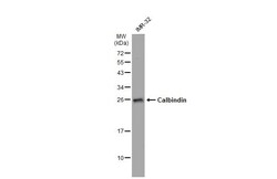
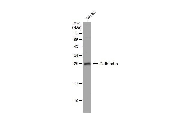
- Western blot analysis of Calbindin D28K was performed by separating 30 µg of IMr-32 whole cell extracts by 12% SDS-PAGE. Proteins were transferred to a membrane and probed with a Calbindin D28K Polyclonal Antibody (Product # PA5-85669) at a dilution of 1:500. The HRP-conjugated anti-rabbit IgG antibody was used to detect the primary antibody.
- Submitted by
- Invitrogen Antibodies (provider)
- Main image
- Experimental details
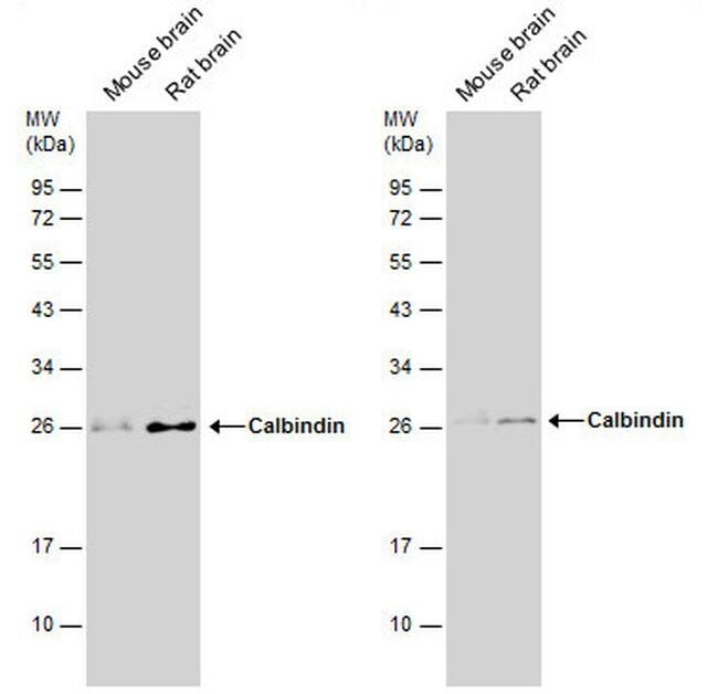
- Western Blot using Calbindin D28K Polyclonal Antibody (Product # PA5-85669). Various tissue extracts (50 µg) were separated by 12% SDS-PAGE, and the membrane was blotted with CALB1antibody Calbindin D28K Polyclonal Antibody (Product # PA5-85669) diluted at 1:1,000. The HRP-conjugated anti-rabbit IgG antibody was used to detect the primary antibody.
Supportive validation
- Submitted by
- Invitrogen Antibodies (provider)
- Main image

- Experimental details
- Immunocytochemistry-Immunofluorescence analysis of Calbindin D28K was performed in DIV9 rat E18 primary cortical neuron cells fixed in 4% paraformaldehyde at RT for 15 min. Green: Calbindin D28K Polyclonal Antibody (Product # PA5 85669) diluted at 1:500. Red: beta Tubulin 3/ Tuj1, stained by beta Tubulin 3/ Tuj1 antibody. Blue: Fluoroshield with DAPI.

Supportive validation
- Submitted by
- Invitrogen Antibodies (provider)
- Main image
- Experimental details
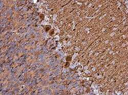
- Calbindin D28K Polyclonal Antibody detects Calbindin protein at cytoplasm in rat brain by immunohistochemical analysis. Sample: Paraffin-embedded rat brain. Calbindin D28K Polyclonal Antibody (Product # PA5-85669) diluted at 1:1,000. Antigen Retrieval: Citrate buffer, pH 6.0, 15 min.

- Submitted by
- Invitrogen Antibodies (provider)
- Main image
- Experimental details
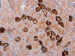
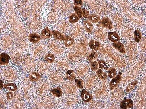
- Calbindin D28K Polyclonal Antibody detects Calbindin protein at cytoplasm in mouse kidney by immunohistochemical analysis. Sample: Paraffin-embedded mouse kidney. Calbindin D28K Polyclonal Antibody (Product # PA5-85669) diluted at 1:1,000. Antigen Retrieval: Citrate buffer, pH 6.0, 15 min.
- Submitted by
- Invitrogen Antibodies (provider)
- Main image

- Experimental details
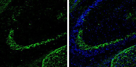
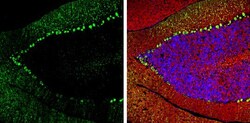
- Calbindin D28K Polyclonal Antibody (Product # PA5-85669) detects Calbindin protein expression by immunohistochemical analysis. Sample: Frozen-sectioned adult mouse hippocampus. Green: Calbindin protein stained by Calbindin D28K Polyclonal Antibody diluted at 1:250. Blue: Fluoroshield with DAPI.
- Submitted by
- Invitrogen Antibodies (provider)
- Main image
- Experimental details
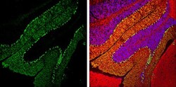
- Calbindin D28K Polyclonal Antibody detects Calbindin protein expression by immunohistochemical analysis. Sample: Frozen-sectioned adult mouse cerebellum. Green: Calbindin protein stained by Calbindin D28K Polyclonal Antibody (Product # PA5-85669) diluted at 1:250. Red: beta Tubulin 3/ TUJ1, stained by beta Tubulin 3/ TUJ1 antibody [GT11710] diluted at 1:500. Blue: Fluoroshield with DAPI .

- Submitted by
- Invitrogen Antibodies (provider)
- Main image
- Experimental details
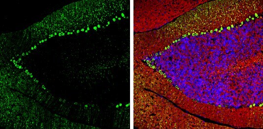
- Calbindin D28K Polyclonal Antibody detects Calbindin protein expression by immunohistochemical analysis. Sample: Frozen-sectioned adult mouse cerebellum. Green: Calbindin protein stained by Calbindin D28K Polyclonal Antibody (Product # PA5-85669) diluted at 1:250. Red: beta Tubulin 3/ TUJ1, stained by beta Tubulin 3/ TUJ1 antibody [GT11710] diluted at 1:500. Blue: Fluoroshield with DAPI .
- Submitted by
- Invitrogen Antibodies (provider)
- Main image
- Experimental details
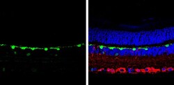
- Immunohistochemistry (Paraffin) analysis of Calbindin D28K was performed in paraffin-embedded mouse retina tissue. Green: Calbindin D28K stained by Calbindin D28K Polyclonal Antibody (Product # PA5-85669) at a dilution of 1:250. Red: beta Tubulin 3/ Tuj1, a marker, stained by beta Tubulin 3/ Tuj1 antibody. Blue: Fluoroshield with DAPI. Antigen Retrieval: Citrate buffer, pH 6.0, 15 min.

Supportive validation
- Submitted by
- Invitrogen Antibodies (provider)
- Main image
- Experimental details
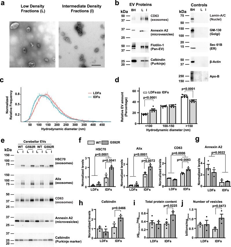
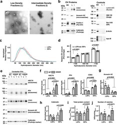
- Fig. 9 The Kv3.3 G592R mutation promotes release of exosomes into the extracellular space. a Representative transmission electron microscopy photomicrographs of low density fraction (LDF) and intermediate-density fraction (IDF) extracellular vesicles (EVs) isolated from the cerebellum of a wild type mouse. Scale bar: 500 nm. Images are representative of ten images taken of each of the sets analyzed. b Representative Western blot analyses of LDF EVs (L) and IDF EVs (I) compared to the cerebellar homogenate (BH). c Nanotrack analysis (NTA) of the hydrodynamic diameter of the EVs found in LDFs and IDFs. The curve is normalized to the mode of each distribution (five independent isolations. d Quantification of particles in LDFs and IDFs by NTA that are found within the size bins shown in the graph. Data are plotted as percentage of total number of EVs. Five independent isolations. e Representative Western blot analyses of EVs isolated from cerebella of wild type and G592R Kv3.3 mutant mice. LDFs (L) and IDFs (I) were isolated from cerebella of two mice of the same genotype and these were combined and tested for the markers shown. Two independent experiments are shown in the blot. f - h quantification of data in e . The densitometric quantification was performed on three independent experiments, representative of six mice per genotype. BCA protein assay ( i ) and total particle count as estimated by NTA ( j ) of LDFs and IDFs in wild type and G592R Kv3.3 mice. Values were normalize