Explore
 Validate
 Learn
Learn Western blot
Antibody data
- Antibody Data
- Antigen structure
- References [1]
- Comments [0]
- Validations
- Western blot [1]
- Immunohistochemistry [1]
- Other assay [1]
Submit
Validation data
Reference
Comment
Report error
- Product number
- 700655 - Provider product page

- Provider
- Invitrogen Antibodies
- Product name
- TARC Recombinant Rabbit Monoclonal Antibody (B22H33L5)
- Antibody type
- Monoclonal
- Antigen
- Recombinant full-length protein
- Description
- Intact IgG appears on a non-reducing gel as ~150 kDa band and upon reduction generating a ~25 kDa light chain band and a ~50 kDa heavy chain.
- Antibody clone number
- B22H33L5
- Concentration
- 0.5 mg/mL
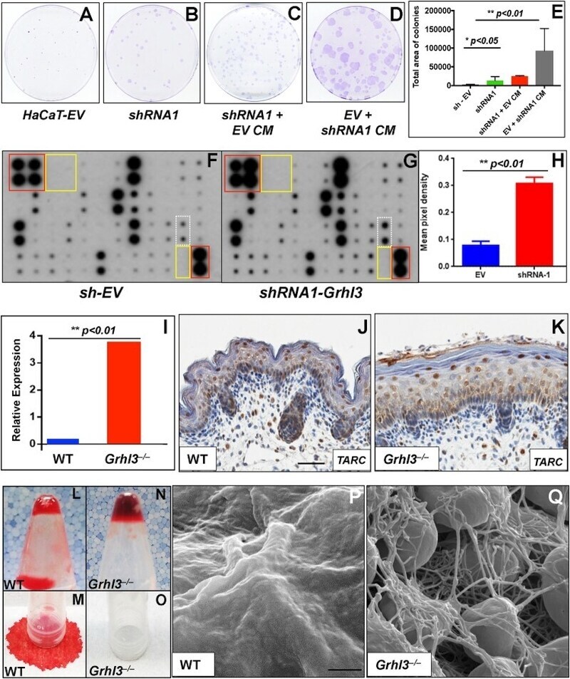
Submitted references Loss of GRHL3 leads to TARC/CCL17-mediated keratinocyte proliferation in the epidermis.
Goldie SJ, Cottle DL, Tan FH, Roslan S, Srivastava S, Brady R, Partridge DD, Auden A, Smyth IM, Jane SM, Dworkin S, Darido C
Cell death & disease 2018 Oct 19;9(11):1072
Cell death & disease 2018 Oct 19;9(11):1072
No comments: Submit comment
Supportive validation
- Submitted by
- Invitrogen Antibodies (provider)
- Main image

- Experimental details
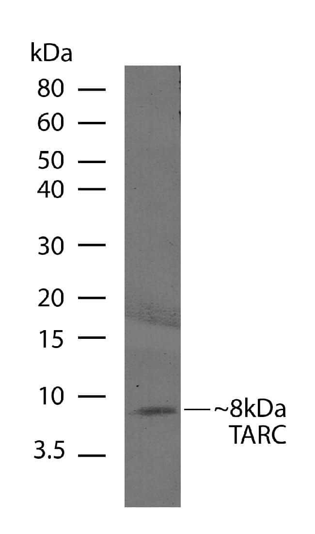
- Western blot analysis of recombinant TARC (5 ng) using a TARC recombinant rabbit monoclonal antibody (Product # 700655) at a dilution of 2.5 µg/mL. NBT/BCIP was used as the substrate (Product # WB7105).
Supportive validation
- Submitted by
- Invitrogen Antibodies (provider)
- Main image
- Experimental details
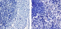
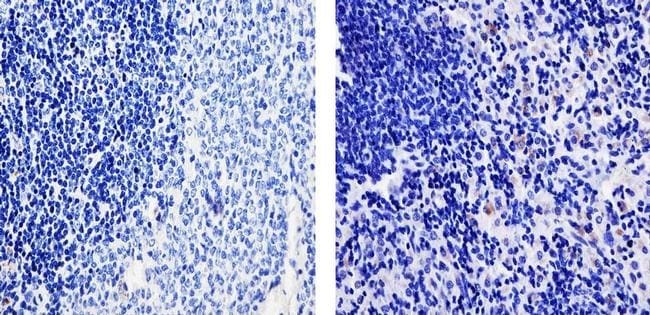
- Immunohistochemistry analysis of TARC showing weak staining in the cytoplasm paraffin-embedded mouse spleen tissue (right) compared to a negative control without primary antibody (left). To expose target proteins, antigen retrieval was performed using 10mM sodium citrate (pH 6.0), microwaved for 8-15 min. Following antigen retrieval, tissues were blocked in 3% H2O2-methanol for 15 min at room temperature, washed with ddH2O and PBS, and then probed with a TARC Recombinant Rabbit Monoclonal Antibody (Product # 700655) diluted in 3% BSA-PBS at a dilution of 1:20 for 1 hour at 37ºC in a humidified chamber. Tissues were washed extensively in PBST and detection was performed using an HRP-conjugated secondary antibody followed by colorimetric detection using a DAB kit. Tissues were counterstained with hematoxylin and dehydrated with ethanol and xylene to prep for mounting.
Supportive validation
- Submitted by
- Invitrogen Antibodies (provider)
- Main image

- Experimental details
- Fig. 1 Loss of GRHL3 leads to increased keratinocyte cell proliferation and elevated TARC expression. KD of GRHL3 via shRNA ( shRNA1-GRHL3 ) leads to increased colony size ( a, b ), relative to transduction with empty vector ( sh-EV ), albeit not increased colony number (data not shown) in the human epidermal keratinocyte cell line HaCaT. Conditioned medium (CM) from EV cultures did not stimulate growth of shRNA1 cells ( c ). However, CM from shRNA1 cultures stimulated significant growth of EV colonies ( d ), a finding confirmed by quantitation of total colony area per plate ( e ). Analysis of cytokine activity in CM collected from HaCaT cells transduced with sh-EV ( f ) or shRNA1-GRHL3 ( g ) shows that the only cytokine that is significantly overexpressed (when quantitated by densitometric scanning; h ) following GRHL3 KD is TARC (dotted white box). Positive (red boxes) and negative controls (yellow boxes) are also shown. Q-RT-PCR quantitation of mRNA expression ( i ) of E18.5 back skin from WT and Grhl3 -/- embryos shows that TARC is significantly elevated in the skin of Grhl3 -/- embryos. Immunohistochemical analysis ( j , k ) shows predominant TARC expression in the nuclei of keratinocytes near or in the granular layer. Weaker basal TARC is also detected. Scale bars correspond to 50 mum. When WT embryonic skin is placed in a microtube with WT adult blood, ( l , m ), no clotting occurs, indicative of an absence of soluble clotting factors in the blo