Explore
 Validate
 Learn
Learn Western blot
 ELISA
ELISAAntibody data
- Antibody Data
- Antigen structure
- References [0]
- Comments [0]
- Validations
- Western blot [1]
- Immunohistochemistry [1]
Submit
Validation data
Reference
Comment
Report error
- Product number
- R1541P - Provider product page

- Provider
- Acris Antibodies GmbH
- Proper citation
- Acris Antibodies GmbH Cat#R1541P, RRID:AB_1004839
- Product name
- anti MAD2L2
- Antibody type
- Polyclonal
- Antigen
- This affinity purified antibody was prepared from whole rabbit serum produced by repeated immunizations with a synthetic peptide corresponding to aa 3-14 of Human MAD2L2.
- Reactivity
- Human
- Host
- Rabbit
- Vial size
- 0.1 mg
- Concentration
- 1.3 mg/ml (by UV absorbance at 280 nm)
No comments: Submit comment
Supportive validation
- Submitted by
- Acris Antibodies GmbH (provider)
- Main image
- Experimental details
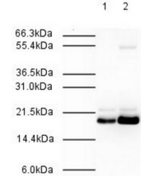
- R1541P MAD2L2 antibody was used at a 1/500 dilution to detect Human MAD2L2 by Western blot. Both HeLa whole cell lysate (Lane 1) and nuclear lysate (Lane 2) were probed using this antibody. This antibody clearly detects a ~20 kDa band corresponding to Human MAD2L2 (predicted molecular weight is 24 kDa). Approximately 20 µg of each lysate was loaded on a 10% SDS-PAGE. Primary antibody was reacted with the membrane at room temperature for 1 h. After subsequent washing, a 1/2,000 dilution of HRP conjugated Goat-anti-Rabbit IgG was used for visualization. Exposure time was 30 sec.

Supportive validation
- Submitted by
- Acris Antibodies GmbH (provider)
- Main image

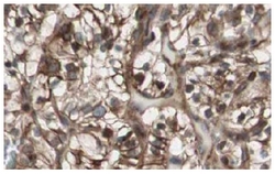
- Experimental details
- R1541P MAD2L2 antibody shows strong nuclear and cytoplasmic staining of tumor cells in cancerous Human kidney tissue. Tissue was formalin-fixed and paraffin embedded. Brown color indicates presence of protein, blue color shows cell nuclei.