 Explore
 Validate
 Learn
Learn Western blot
 Immunoprecipitation
Antibody data
- Antibody Data
- Antigen structure
- References [0]
- Comments [0]
- Validations
- Western blot [3]
- Immunocytochemistry [3]
- Immunohistochemistry [2]
Submit
Validation data
Reference
Comment
Report error
- Product number
- MA5-34803 - Provider product page

- Provider
- Invitrogen Antibodies
- Product name
- CtBP1 Recombinant Rabbit Monoclonal Antibody (JG39-73)
- Antibody type
- Monoclonal
- Antigen
- Recombinant full-length protein
- Description
- Positive Control: PC-3M, rat kidney tissue, human colon tissue, A549.
- Reactivity
- Human, Mouse, Rat
- Host
- Rabbit
- Isotype
- IgG
- Antibody clone number
- JG39-73
- Vial size
- 100 µL
- Concentration
- 1 mg/mL
- Storage
- -20° C, Avoid Freeze/Thaw Cycles, store in dark
No comments: Submit comment
Supportive validation
- Submitted by
- Invitrogen Antibodies (provider)
- Main image

- Experimental details
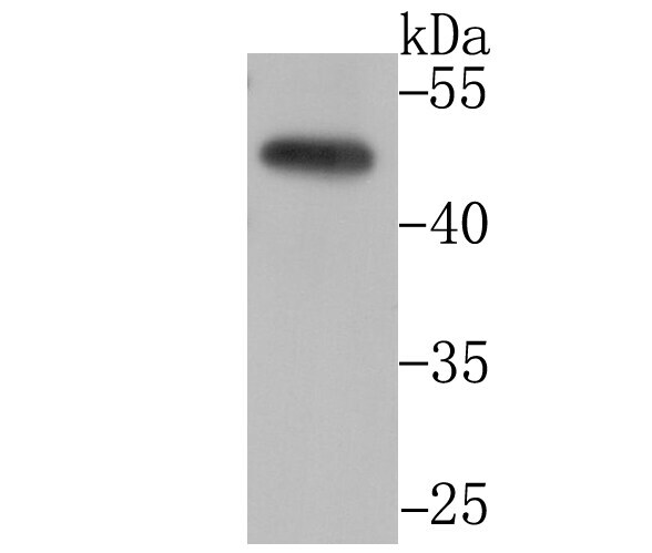
- Western blot analysis of CtBP1 in PC-3M cell lysate. Samples were incubated with CtBP1 monoclonal antibody (Product # MA5-34803), at a dilution of 1:1000.
- Submitted by
- Invitrogen Antibodies (provider)
- Main image

- Experimental details
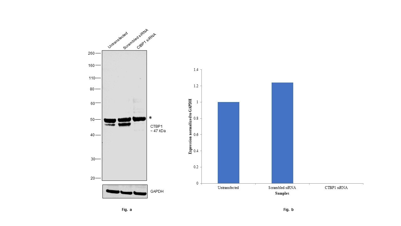
- Knockdown of CtBP1 was achieved by transfecting HEK-293 with CtBP1 specific siRNAs (Silencer® select Product # S3698, S3699). Western blot analysis (Fig. a) was performed using whole cell extracts from the CtBP1 knockdown cells (lane 3), non-targeting scrambled siRNA transfected cells (lane 2) and untransfected cells (lane 1). The blot was probed with CtBP1 Recombinant Rabbit Monoclonal Antibody (JG39-73) (Product # MA5-34803, 1:1000) and Goat anti-Rabbit IgG (H+L) Superclonal™ Recombinant Secondary Antibody, HRP (Product # A27036, 1:20,000). Densitometric analysis of this western blot is shown in histogram (Fig. b). Decrease in signal upon siRNA mediated knock down confirms that antibody is specific to CtBP1. The other band could be an isoform of CtBP1 (*). CtBP2 knockdown was also performed and the blot was probed with CtBP1 Recombinant Rabbit Monoclonal Antibody (JG39-73) (Product # MA5-34803, 1:1000), and showed no knockdown (data not shown). This ensures the antibody is specific to CtBP1.
- Submitted by
- Invitrogen Antibodies (provider)
- Main image
- Experimental details
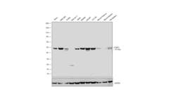
- Western blot was performed using CtBP1 Recombinant Rabbit Monoclonal Antibody (JG39-73) (Product # MA5-34803) and a ~47 kDa band corresponding to CtBP1 was observed across all cell lines and tissues. Whole cell extracts (30 µg lysate) of HeLa (Lane 1), HEK-293 (Lane 2), K-562 (Lane 3), Kasumi-1 (Lane 4), A549 (Lane 5), SW480 (Lane 6), LNCaP (Lane 7), U-2 OS (Lane 8), Mouse Thymus (Lane 9), Mouse Spleen (Lane 10), Rat Spleen (Lane 11) were electrophoresed using NuPAGE™ 4-12% Bis-Tris Protein Gel (Product # NP0322BOX), 12 well. Resolved proteins were then transferred onto a nitrocellulose membrane (Product # IB23001) by iBlot® 2 Dry Blotting System (Product # IB21001). The blot was probed with the primary antibody (1:1000) and detected by chemiluminescence with Goat anti-Rabbit IgG (H+L) Superclonal™ Recombinant Secondary Antibody, HRP (Product # A27036, 1:20,000) using the iBright™ FL1500 Imaging System (Product # A44115). Chemiluminescent detection was performed using SuperSignal™ West Pico PLUS Chemiluminescent Substrate (Product # 34580).

Supportive validation
- Submitted by
- Invitrogen Antibodies (provider)
- Main image
- Experimental details
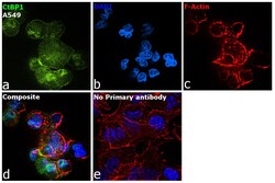
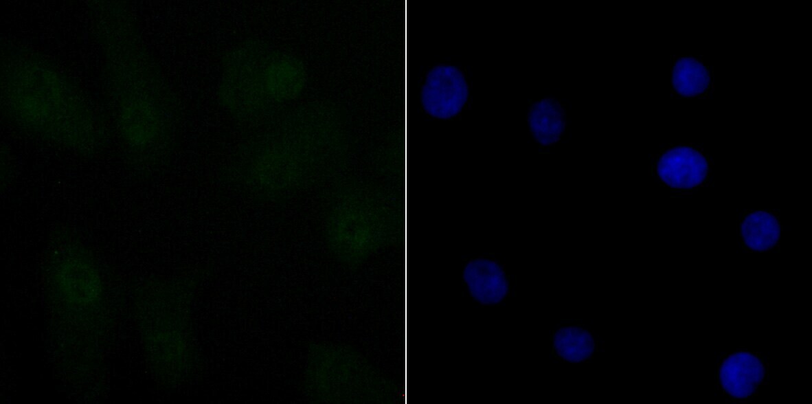
- Immunofluorescence analysis of CtBP1 was performed using 70% confluent log phase A549 cells. The cells were fixed with 4% paraformaldehyde for 10 minutes, permeabilized with 0.1% Triton™ X-100 for 10 minutes, and blocked with 2% BSA for 45 minutes at room temperature. The cells were labeled with CtBP1 Recombinant Rabbit Monoclonal Antibody (JG39-73) (Product # MA5-34803) at 1:100 in 0.1% BSA, incubated at 4 degree celsius overnight and then labeled with Donkey anti-Rabbit IgG (H+L) Highly Cross-Adsorbed Secondary Antibody, Alexa Fluor™ Plus 488 (Product # A32790, 1:2000), for 45 minutes at room temperature (Panel a: Green). Nuclei (Panel b:Blue) were stained with ProLong™ Diamond Antifade Mountant with DAPI (Product # P36962). F-actin (Panel c: Red) was stained with Rhodamine Phalloidin (Product # R415, 1:300). Panel d represents the merged image showing nucleus and cytoplasmic localization. Panel e represents control cells with no primary antibody to assess background. The images were captured at 60X magnification.

- Submitted by
- Invitrogen Antibodies (provider)
- Main image

- Experimental details
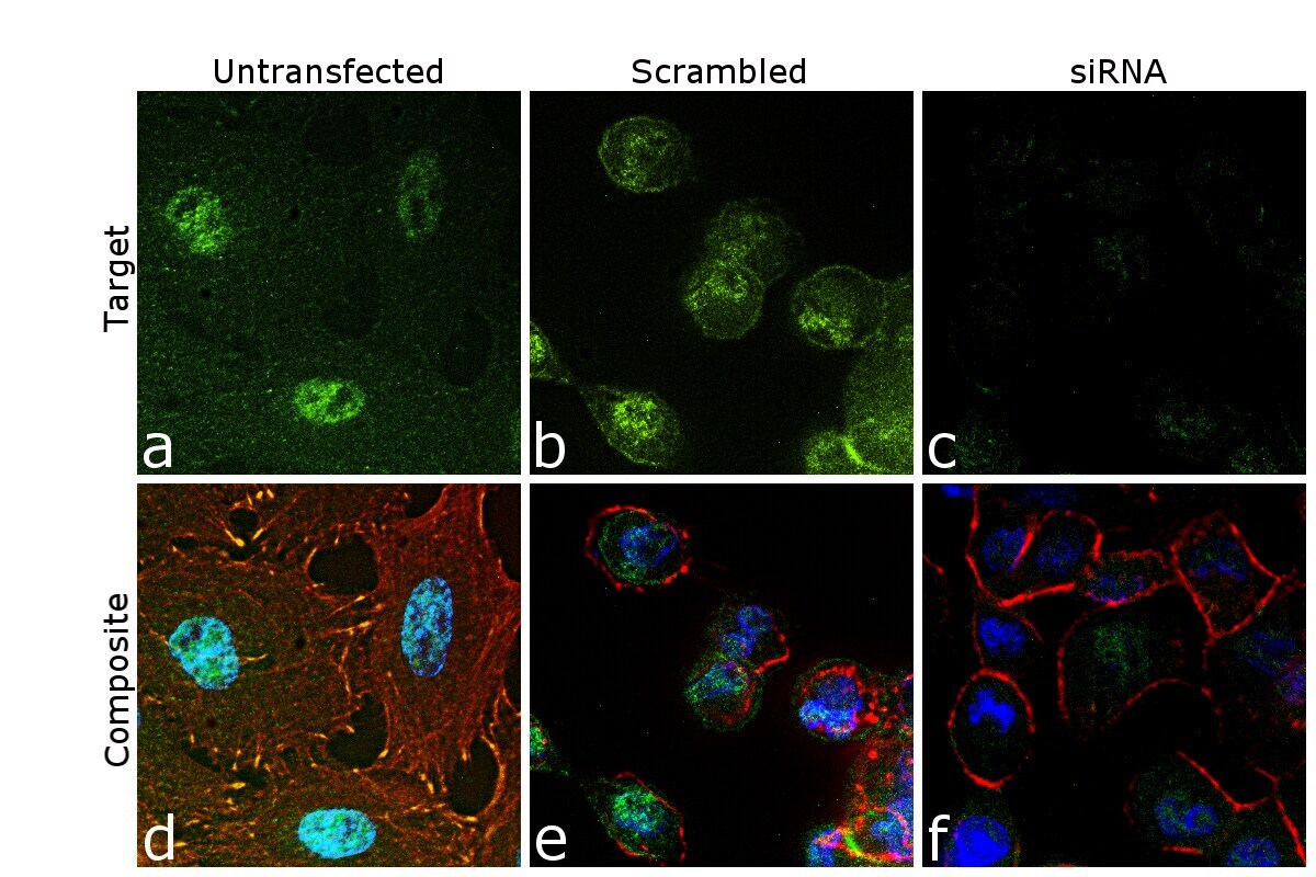
- Knockdown of CtBP1 was achieved by transfecting A549 cells with CtBP1 specific siRNA (Silencer® select Product # S3698, S3699). Immunofluorescence analysis was performed on untransfected A549 cells (panel a,d), transfected with non-specific scrambled siRNA (panels b,e) and transfected with CtBP1 specific siRNA (panel c,f). Cells were fixed, permeabilized, and labelled with CtBP1 Recombinant Rabbit Monoclonal Antibody (JG39-73) (Product # MA5-34803, 1:100) followed by Donkey anti-Rabbit IgG (H+L) Highly Cross-Adsorbed Secondary Antibody, Alexa Fluor™ Plus 488 (Product # A32790, 1:2000). Nuclei (blue) were stained using ProLong™ Diamond Antifade Mountant with DAPI (Product # P36962), and Rhodamine Phalloidin (Product # R415, 1:300) was used for cytoskeletal F-actin (Red) staining , and specific signal was observed upon siRNA mediated knockdown (panel c,f) confirming specificity of the antibody to CtBP1 (Green). The Images were captured at 60X magnification.
- Submitted by
- Invitrogen Antibodies (provider)
- Main image
- Experimental details
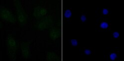
- Immunofluorescent analysis of CtBP1 in A549 cells (green). Samples were fixed in paraformaldehyde and permeabilised with 0.25% Triton X100/PBS, incubated with CtBP1 monoclonal antibody (Product # MA5-34803), followed by DAPI (blue).
Supportive validation
- Submitted by
- Invitrogen Antibodies (provider)
- Main image
- Experimental details
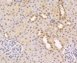
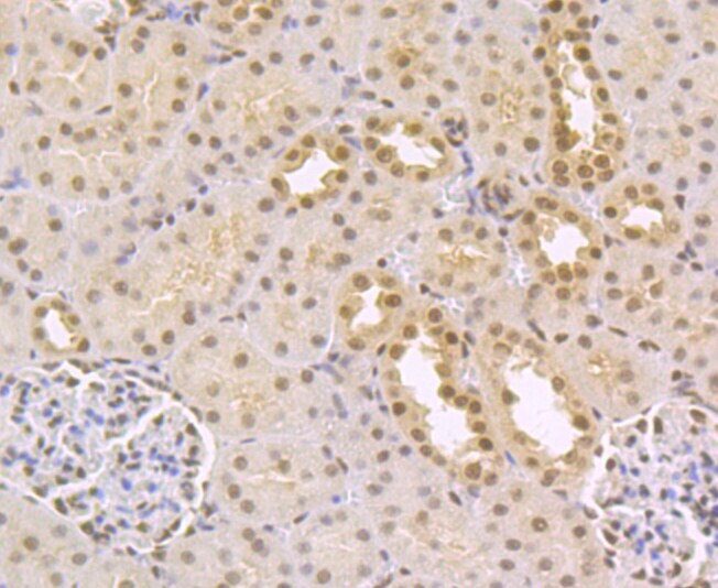
- Immunohistochemistry analysis of CtBP1 in paraffin-embedded rat kidney tissue. Samples were incubated with CtBP1 monoclonal antibody (Product # MA5-34803), and followed by hematoxylin.
- Submitted by
- Invitrogen Antibodies (provider)
- Main image
- Experimental details
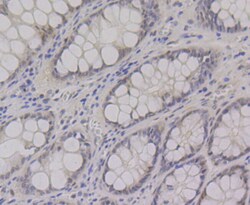
- Immunohistochemistry analysis of CtBP1 in paraffin-embedded human colon tissue. Samples were incubated with CtBP1 monoclonal antibody (Product # MA5-34803), and followed by hematoxylin.
