Explore
 Validate
 Learn
Learn Western blot
Antibody data
- Antibody Data
- Antigen structure
- References [0]
- Comments [0]
- Validations
- Western blot [4]
- Immunocytochemistry [3]
- Immunohistochemistry [1]
Submit
Validation data
Reference
Comment
Report error
- Product number
- PA5-87361 - Provider product page

- Provider
- Invitrogen Antibodies
- Product name
- RACK1 Polyclonal Antibody
- Antibody type
- Polyclonal
- Antigen
- Recombinant full-length protein
- Description
- Immunogen sequence: MILSASRDKT IIMWKLTRDE TNYGIPQRAL RGHSHFVSDV VISSDGQFAL SGSWDGTLRL WDLTTGTTTR RFVGHTKDVL SVAFSSDNRQ IVSGSRDKTI KLWNTLGVCK YTVQDESHSE WVSCVRFSPN SSNPIIVSCG WDKLVKVWNL ANCKLKTNHI GHTGYLNTVT VSPDGSLCAS GGKDGQAMLW DLNEGKHLYT LDGGDIINAL CFSPNRYWLC AATGPSIKIW DLEGKIIVDE LKQEVISTSS KAEPPQCTSL A; Positive Samples: MCF-7, 293T, Mouse spleen; Cellular Location: Cell membrane, Cell projection, Cytoplasm, Nucleus, Perikaryon, Peripheral membrane protein, cytoskeleton, dendrite, perinuclear region, phagocytic cup
- Reactivity
- Human, Mouse
- Host
- Rabbit
- Isotype
- IgG
- Vial size
- 100 µL
- Concentration
- 0.16 mg/mL
- Storage
- -20° C, Avoid Freeze/Thaw Cycles
No comments: Submit comment
Supportive validation
- Submitted by
- Invitrogen Antibodies (provider)
- Main image
- Experimental details
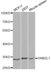
- Western blot analysis of extracts of various cell lines, using GNB2L1 Polyclonal antibody (Product # PA5-87361) at 1:1000 dilution. Secondary antibody: HRP Goat Anti-Rabbit IgG (H+L) at 1:10000 dilution. Lysates/proteins: 25ug per lane. Blocking buffer: 3% nonfat dry milk in TBST.

- Submitted by
- Invitrogen Antibodies (provider)
- Main image
- Experimental details
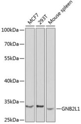
- Western Blot analysis of RACK1 in extracts of various cell lines using RACK1 Polyclonal Antibody (Product # PA5-87361) at a dilution of 1:1000. A HRP Goat Anti-Rabbit IgG (H+L) secondary antibody was used at a dilution of 1:10,000. Lysates/proteins: 25 µg per lane. Blocking buffer: 3% nonfat dry milk in TBST.

- Submitted by
- Invitrogen Antibodies (provider)
- Main image
- Experimental details
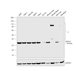
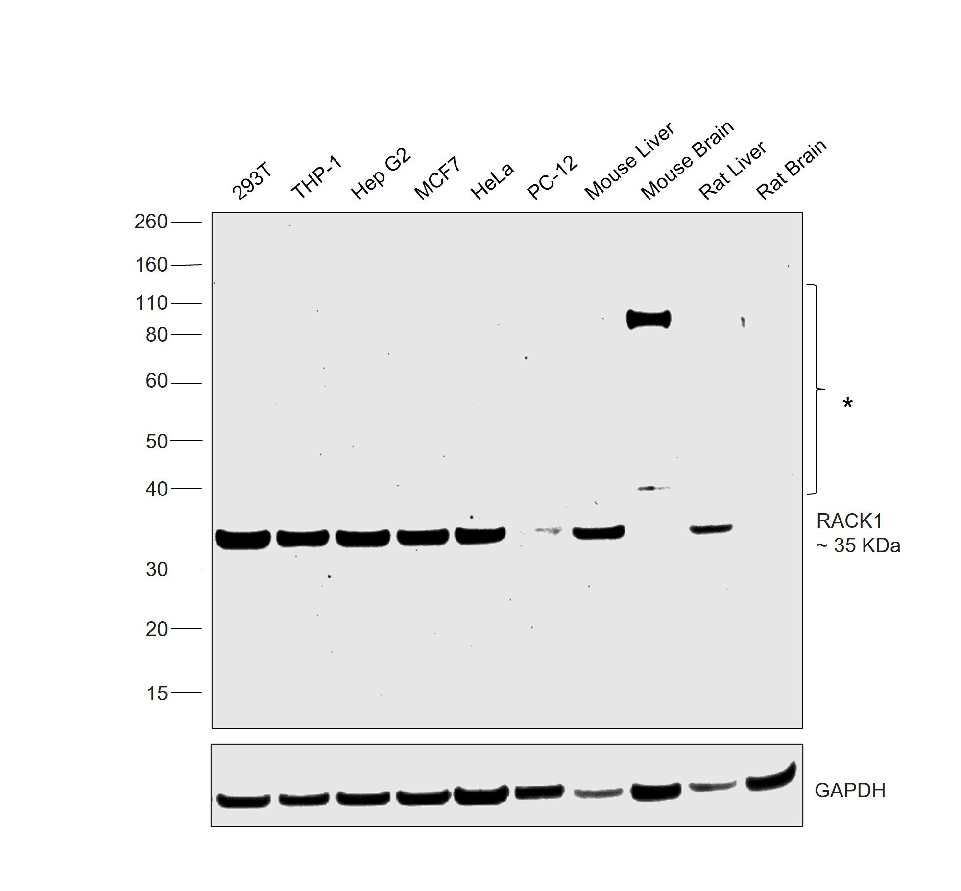
- Western blot was performed using Anti-RACK1 Polyclonal Antibody (Product # PA5-87361) and a 35 kDa band corresponding to RACK1 was observed across across cell lines tested. Whole cell extracts (30 µg lysate) of HEK-293 (Lane 1), THP-1 (Lane 2), Hep G2 (Lane 3), MCF7 (Lane 4), HeLa (Lane 5), PC-12 (Lane 6), Mouse Liver (Lane 7), Mouse Brain (Lane 8), Rat Liver (Lane 9) and Rat Brain (Lane 10) were electrophoresed using NuPAGE™ 4-12% Bis-Tris Protein Gel (Product # NP0322BOX). Resolved proteins were then transferred onto a nitrocellulose membrane (Product # IB23001) by iBlot® 2 Dry Blotting System (Product # IB21001). The blot was probed with the primary antibody (1:1000 dilution) and detected by chemiluminescence with Goat anti-Rabbit IgG (H+L) Superclonal™ Recombinant Secondary Antibody, HRP (Product # A27036,1:20000) using the iBright™ FL1500 Imaging System (Product # A44115). Chemiluminescent detection was performed using SuperSignal™ West Pico PLUS Chemiluminescent Substrate (Product # 34580).
- Submitted by
- Invitrogen Antibodies (provider)
- Main image
- Experimental details
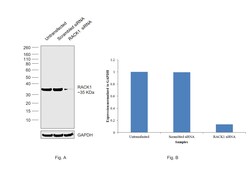
- Knockdown of RACK1 was achieved by transfecting HeLa with RACK1 specific siRNAs (Silencer® select Product # s20342, s20340). Western blot analysis (Fig. a) was performed using Whole cell extracts from the RACK1 knockdown cells (lane 3), non-targeting scrambled siRNA transfected cells (lane 2) and untransfected cells (lane 1). The blot was probed with RACK1 Polyclonal Antibody (Product # PA5-87361, 1:1000 dilution ) and Goat anti-Rabbit IgG (H+L) Superclonal™ Recombinant Secondary Antibody, HRP (Product # A27036, 1:20,000 dilution). Densitometric analysis of this western blot is shown in histogram (Fig. b). Decrease in signal upon siRNA mediated knock down confirms that antibody is specific to RACK1.

Supportive validation
- Submitted by
- Invitrogen Antibodies (provider)
- Main image
- Experimental details
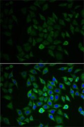
- Immunocytochemistry-Immunofluorescence analysis of RACK1 was performed in A-549 cells using RACK1 Polyclonal Antibody (Product # PA5-87361). Blue: DAPI for nuclear staining.

- Submitted by
- Invitrogen Antibodies (provider)
- Main image
- Experimental details
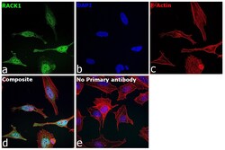
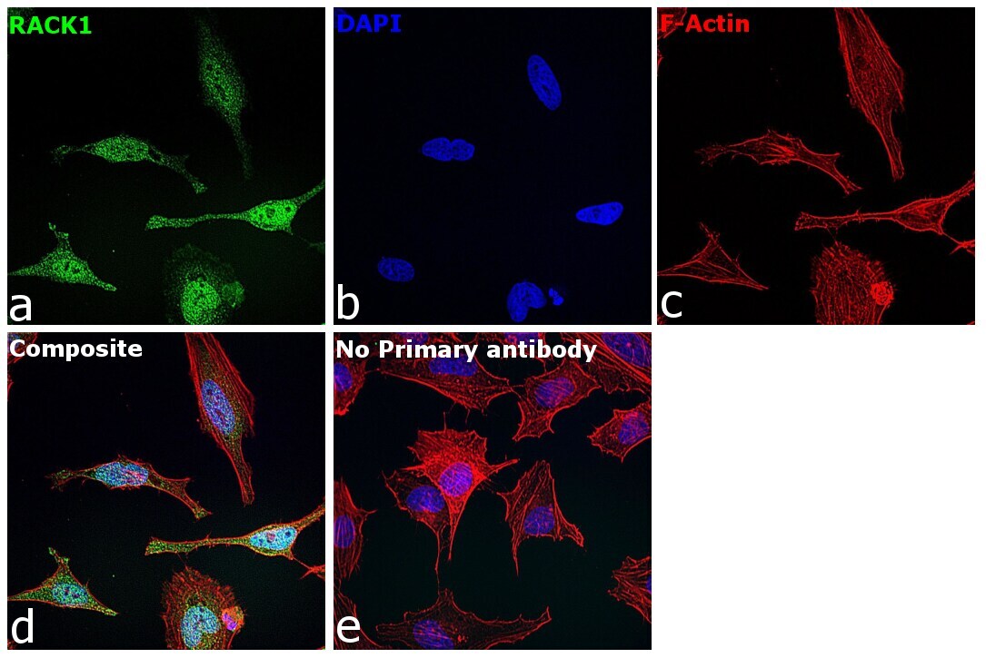
- Immunofluorescence analysis of RACK1 was performed using 70% confluent log phase HeLa cells. The cells were fixed with 4% paraformaldehyde for 10 minutes, permeabilized with 0.1% Triton™ X-100 for 10 minutes, and blocked with 2% BSA for 1 hour at room temperature. The cells were labeled with RACK1 Polyclonal Antibody (Product # PA5-87361) at 1:100 dilution in 0.1% BSA, incubated at 4 degree celsius overnight and then labeled with Goat anti-Rabbit IgG (H+L) Superclonal™ Recombinant Secondary Antibody, Alexa Fluor® 488 conjugate (Product # A27034), (1:2000), for 45 minutes at room temperature (Panel a: Green). Nuclei (Panel b:Blue) were stained with Hoechst 33342 (Product # H1399). F-actin (Panel c: Red) was stained with Alexa Fluor™ Plus 647 Phalloidin (Product # A30107, 1:2000 dilution). Panel d represents the merged image showing nucleus, cytoplasm localization. Panel e represents control cells with no primary antibody to assess background. The images were captured at 40X magnification. The images were captured at 40X magnification in CellInsight CX7 LZR High-Content Screening (HCS) Platform (Product # CX7A1110LZR) and externally deconvoluted (D.Sage et al./Methods 115 (2017) 28–41.
- Submitted by
- Invitrogen Antibodies (provider)
- Main image

- Experimental details
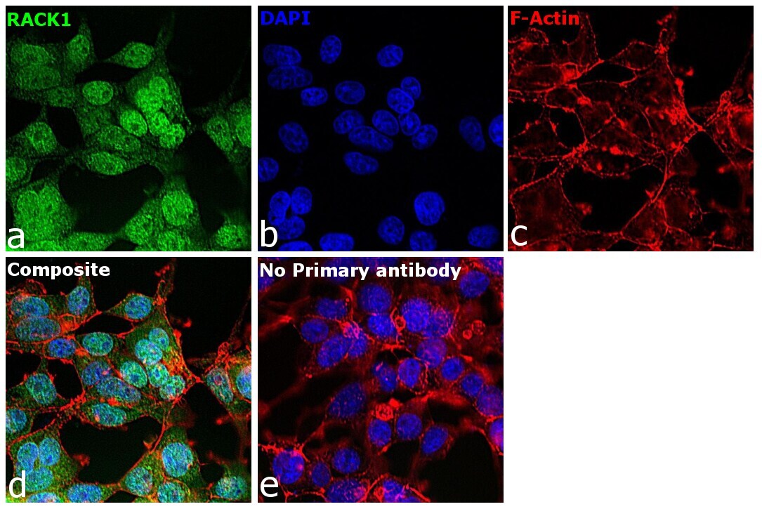
- Immunofluorescence analysis of RACK1 was performed using 70% confluent log phase HEK-293 cells. The cells were fixed with 4% paraformaldehyde for 10 minutes, permeabilized with 0.1% Triton™ X-100 for 10 minutes, and blocked with 2% BSA for 1 hour at room temperature. The cells were labeled with RACK1 Polyclonal Antibody (Product # PA5-87361) at 1:100 dilution in 0.1% BSA, incubated at 4 degree celsius overnight and then labeled with Goat anti-Rabbit IgG (H+L) Superclonal™ Recombinant Secondary Antibody, Alexa Fluor® 488 conjugate (Product # A27034), (1:2000), for 45 minutes at room temperature (Panel a: Green). Nuclei (Panel b:Blue) were stained with Hoechst 33342 (Product # H1399). F-actin (Panel c: Red) was stained with Alexa Fluor™ Plus 647 Phalloidin (Product # A30107, 1:2000 dilution). Panel d represents the merged image showing nucleus, cytoplasm localization. Panel e represents control cells with no primary antibody to assess background. The images were captured at 40X magnification. The images were captured at 40X magnification in CellInsight CX7 LZR High-Content Screening (HCS) Platform (Product # CX7A1110LZR) and externally deconvoluted (D.Sage et al./Methods 115 (2017) 28–41.
Supportive validation
- Submitted by
- Invitrogen Antibodies (provider)
- Main image

- Experimental details
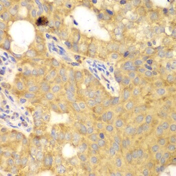
- Immunohistochemistry analysis of RACK1 in paraffin-embedded human oophoroma using RACK1 Polyclonal Antibody (Product # PA5-87361) at a dilution of 1:100.