Explore
 Validate
 Learn
Learn Western blot
 ELISA
ELISAAntibody data
- Antibody Data
- Antigen structure
- References [1]
- Comments [0]
- Validations
- Western blot [2]
- Flow cytometry [1]
Submit
Validation data
Reference
Comment
Report error
- Product number
- LF-MA0015 - Provider product page

- Provider
- Invitrogen Antibodies
- Product name
- Anti-TrxR1 Monoclonal Antibody (19A1)
- Antibody type
- Monoclonal
- Antigen
- Recombinant full-length protein
- Description
- A suggested positive control for this product is HeLa cells.
- Reactivity
- Human
- Host
- Mouse
- Isotype
- IgG
- Antibody clone number
- 19A1
- Vial size
- 100 µL
- Storage
- -20° C, Avoid Freeze/Thaw Cycles
Submitted references Selective up-regulation of human selenoproteins in response to oxidative stress.
Touat-Hamici Z, Legrain Y, Bulteau AL, Chavatte L
The Journal of biological chemistry 2014 May 23;289(21):14750-61
The Journal of biological chemistry 2014 May 23;289(21):14750-61
No comments: Submit comment
Supportive validation
- Submitted by
- Invitrogen Antibodies (provider)
- Main image

- Experimental details
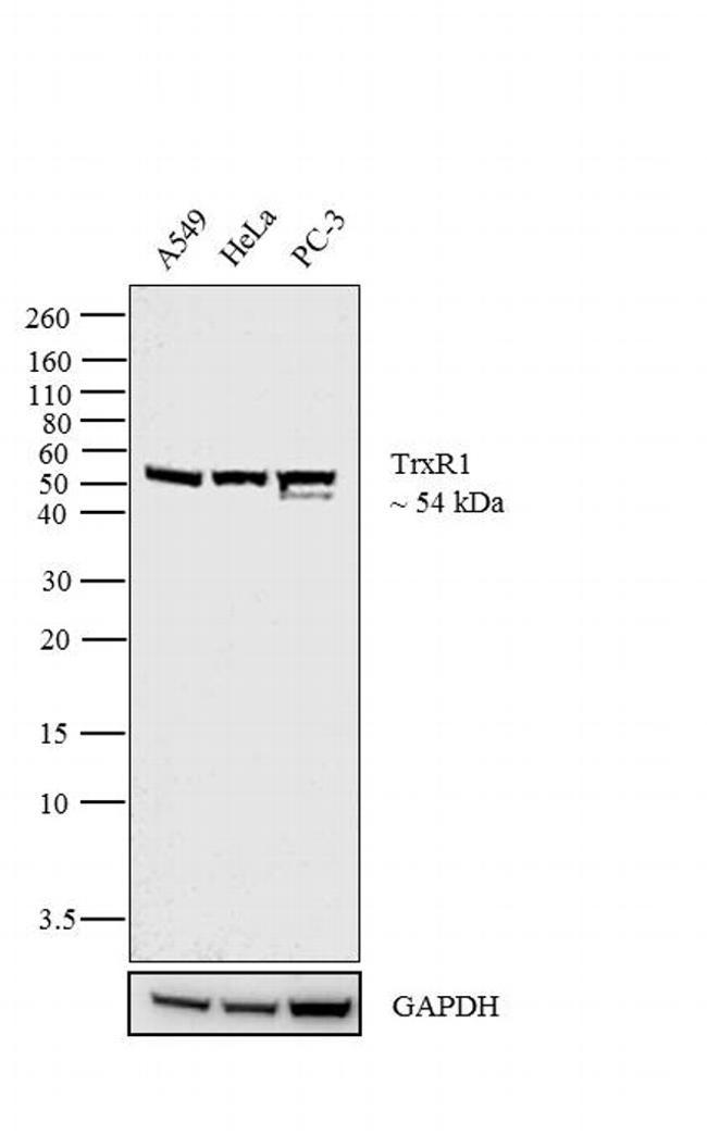
- Western blot analysis was performed on membrane enriched extracts (30 µg lysate) of A549 (Lane 1), HeLa (Lane 2) and PC-3 (Lane 3). The blot was probed with Anti-TrxR1 Mouse Monoclonal Antibody (Product # LF-MA0015, 1:1,000 dilution) and detected by chemiluminescence using Goat anti-Mouse IgG (H+L) Superclonal Secondary Antibody, HRP conjugate (Product # A28177, 0.4 µg/mL, 1:2500 dilution). A 54 kDa band corresponding to TrxR1 was observed cross the cell lines tested. Known quantity of protein samples were electrophoresed using Novex®NuPAGE®4-12 % Bis-Tris gel (Product # NP0321BOX), XCell SureLock Electrophoresis System (Product # EI0002) and Novex® Sharp Pre-Stained Protein Standard (Product # LC5800). Resolved proteins were then transferred onto a nitrocellulose membrane with iBlot® 2 Dry Blotting System (Product # IB21001). The membrane was probed with the relevant primary and secondary Antibody following blocking with 5 % skimmed milk. Chemiluminescent detection was performed using Pierce™ ECL Western Blotting Substrate (Product # 32106).
- Submitted by
- Invitrogen Antibodies (provider)
- Main image
- Experimental details
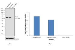
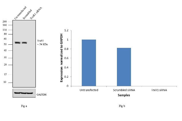
- Knockdown of TrxR1 was achieved by transfecting A549 cells with TrxR1 specific siRNAs (Silencer® select Product # s755). Western blot analysis (Fig a) was performed using whole cell extracts from the TrxR1 knock down cells (lane 3), non-specific scrambled siRNA transfected cells (lane 2) and un-transfected cells (lane 1). The blots were probed with Anti- TrxR1 Mouse monoclonal Antibody (Product # LF-MA0015, 1:500 dilution) and Goat anti-Mouse IgG (H+L) Superclonal™ Secondary Antibody, HRP conjugate (Product # A28177, 0.25 µg/mL, 1:4000 dilution). Densitometric analysis of this western blot is shown in histogram (Fig b). Loss of signal upon siRNA mediated knock down confirms that antibody is specific to TrxR1.
Supportive validation
- Submitted by
- Invitrogen Antibodies (provider)
- Main image

- Experimental details
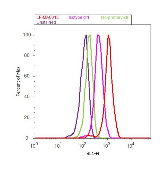
- Flow cytometry analysis of TrxR1 was done on NTERA-2 cells. Cells were fixed with 70% ethanol for 10 minutes, permeabilized with 0.25% Triton™ X-100 for 20 minutes, and blocked with 5% BSA for 30 minutes at room temperature. Cells were labeled with TrxR1 Mouse Monoclonal Antibody (Product # LF-MA0015, red histogram) at 1:20 dilution or a matched isotype control (pink histogram) in 2.5% BSA. After incubation at room temperature for 2 hours, the cells were labeled with Alexa Fluor® 488 Anti-Mouse Secondary Antibody (Product # A-11059) at a dilution of 1:400 for 30 minutes at room temperature. A representative 10,000 cells were acquired and analyzed for each sample using an Attune® Acoustic Focusing Cytometer. The purple histogram represents unstained control cells and the green histogram represents no-primary-antibody control.