Explore
 Validate
 Learn
Learn Western blot
 Immunocytochemistry
Antibody data
- Antibody Data
- Antigen structure
- References [10]
- Comments [0]
- Validations
- Western blot [3]
- Immunohistochemistry [3]
- Other assay [2]
Submit
Validation data
Reference
Comment
Report error
- Product number
- PA5-16754 - Provider product page

- Provider
- Invitrogen Antibodies
- Product name
- Anti-VEGF Polyclonal Antibody
- Antibody type
- Polyclonal
- Antigen
- Synthetic peptide
- Description
- PA5-16754 targets Vascular Endothelial Growth Factor (VEGF) in IF, IHC (P), IP, and WB applications and shows reactivity with Human samples. The PA5-16754 immunogen is a synthetic peptide derived from N-terminal of human VEGF.
- Reactivity
- Human, Mouse
- Host
- Rabbit
- Isotype
- IgG
- Vial size
- 500 µL
- Concentration
- 0.2 mg/mL
- Storage
- 4° C
Submitted references The role of miR-24 as a race related genetic factor in prostate cancer.
Endocardial-to-mesenchymal transformation and mesenchymal cell colonization at the onset of human cardiac valve development.
Deletion of IL-33R attenuates VEGF expression and enhances necrosis in mammary carcinoma.
Combined use of COX-1 and VEGF immunohistochemistry refines the histopathologic prognosis of renal cell carcinoma.
Profilin-1 phosphorylation directs angiocrine expression and glioblastoma progression through HIF-1α accumulation.
Tumor endothelium FasL establishes a selective immune barrier promoting tolerance in tumors.
Aging-related changes of optic nerve of Wistar albino rats.
Castration resistant prostate cancer is associated with increased blood vessel stabilization and elevated levels of VEGF and Ang-2.
Proliferation and maturation of microvessels in arteriovenous malformations--expression patterns of angiogenic and cell cycle-dependent factors.
Inflammation modulates expression of laminin in the central nervous system following ischemic injury.
Hashimoto Y, Shiina M, Kato T, Yamamura S, Tanaka Y, Majid S, Saini S, Shahryari V, Kulkarni P, Dasgupta P, Mitsui Y, Sumida M, Deng G, Tabatabai L, Kumar D, Dahiya R
Oncotarget 2017 Mar 7;8(10):16581-16593
Oncotarget 2017 Mar 7;8(10):16581-16593
Endocardial-to-mesenchymal transformation and mesenchymal cell colonization at the onset of human cardiac valve development.
Monaghan MG, Linneweh M, Liebscher S, Van Handel B, Layland SL, Schenke-Layland K
Development (Cambridge, England) 2016 Feb 1;143(3):473-82
Development (Cambridge, England) 2016 Feb 1;143(3):473-82
Deletion of IL-33R attenuates VEGF expression and enhances necrosis in mammary carcinoma.
Milosavljevic MZ, Jovanovic IP, Pejnovic NN, Mitrovic SL, Arsenijevic NN, Simovic Markovic BJ, Lukic ML
Oncotarget 2016 Apr 5;7(14):18106-15
Oncotarget 2016 Apr 5;7(14):18106-15
Combined use of COX-1 and VEGF immunohistochemistry refines the histopathologic prognosis of renal cell carcinoma.
Osman WM, Youssef NS
International journal of clinical and experimental pathology 2015;8(7):8165-77
International journal of clinical and experimental pathology 2015;8(7):8165-77
Profilin-1 phosphorylation directs angiocrine expression and glioblastoma progression through HIF-1α accumulation.
Fan Y, Potdar AA, Gong Y, Eswarappa SM, Donnola S, Lathia JD, Hambardzumyan D, Rich JN, Fox PL
Nature cell biology 2014 May;16(5):445-56
Nature cell biology 2014 May;16(5):445-56
Tumor endothelium FasL establishes a selective immune barrier promoting tolerance in tumors.
Motz GT, Santoro SP, Wang LP, Garrabrant T, Lastra RR, Hagemann IS, Lal P, Feldman MD, Benencia F, Coukos G
Nature medicine 2014 Jun;20(6):607-15
Nature medicine 2014 Jun;20(6):607-15
Aging-related changes of optic nerve of Wistar albino rats.
El-Sayyad HI, Khalifa SA, El-Sayyad FI, Al-Gebaly AS, El-Mansy AA, Mohammed EA
Age (Dordrecht, Netherlands) 2014 Apr;36(2):519-32
Age (Dordrecht, Netherlands) 2014 Apr;36(2):519-32
Castration resistant prostate cancer is associated with increased blood vessel stabilization and elevated levels of VEGF and Ang-2.
Tomić TT, Gustavsson H, Wang W, Jennbacken K, Welén K, Damber JE
The Prostate 2012 May 15;72(7):705-12
The Prostate 2012 May 15;72(7):705-12
Proliferation and maturation of microvessels in arteriovenous malformations--expression patterns of angiogenic and cell cycle-dependent factors.
Meijer-Jorna LB, van der Loos CM, Teeling P, de Boer OJ, Florquin S, van der Horst CM, van der Wal AC
Journal of cutaneous pathology 2012 Jun;39(6):610-20
Journal of cutaneous pathology 2012 Jun;39(6):610-20
Inflammation modulates expression of laminin in the central nervous system following ischemic injury.
Ji K, Tsirka SE
Journal of neuroinflammation 2012 Jul 3;9:159
Journal of neuroinflammation 2012 Jul 3;9:159
No comments: Submit comment
Supportive validation
- Submitted by
- Invitrogen Antibodies (provider)
- Main image
- Experimental details
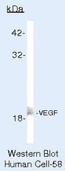
- Western blot of Vascular Endothelial Growth Factor using Vascular Endothelial Growth Factor Polyclonal Antibody (Product # PA5-16754) on VEGF Cells.

- Submitted by
- Invitrogen Antibodies (provider)
- Main image
- Experimental details
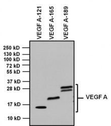
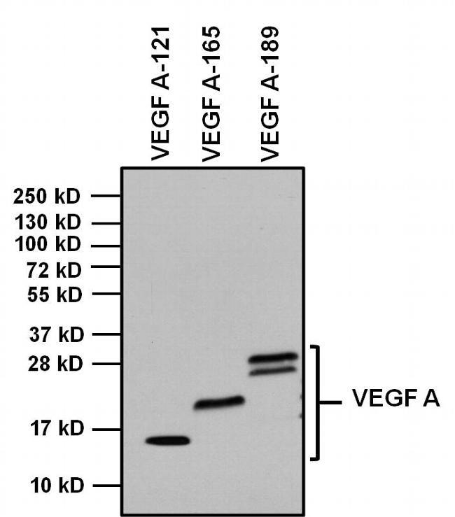
- Western blot analysis of Vascular Endothelial Growth Factor (VEGF) was performed by loading 1 µg of indicated recombinant VEGF isoforms, and 10 µL of PageRuler PlusPrestained Protein Ladder (Product # 26619) per well onto a 4-20% Tris-Glycine polyacrylamide gel. Proteins were transferred to a PVDF membrane (Product # 88518) using the G2 Fast Blotter (Product # 62288) and blocked with 5% Milk/TBST for at least 1 hour at room temperature. VEGF was detected at 14 kD, 19 kD and 22-30 kD using a VEGF polyclonal antibody (Product # PA5-16754) at a dilution of 1:500 in blocking buffer overnight at 4C on a rocking platform, followed by an HRP-conjugated goat anti-rabbit IgG (Fc) secondary antibody (Product # 31463) at a dilution of 1:20,000 for at least 1 hour. Chemiluminescent detection was performed using SuperSignal West Dura (Product # 34076).
- Submitted by
- Invitrogen Antibodies (provider)
- Main image
- Experimental details
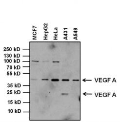
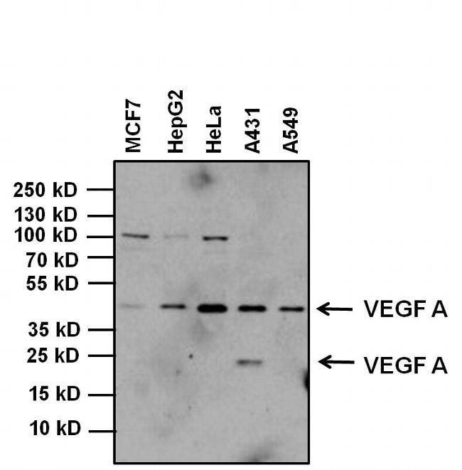
- Western blot analysis of Vascular Endothelial Growth Factor (VEGF) was performed by loading 20 µg of MCF7, HepG2, HeLa, A431, A549 whole cell lysates and 10uL of PageRuler Plus Prestained Protein Ladder (Product # 26619) per well onto a 4-20% Tris-Glycine polyacrylamide gel. Proteins were transferred to a nitrocellulose membrane using the G2 Blotter (Product # 62288), and blocked with 5% milk in TBST for 1 hour at room temperature. VEGF was detected at ~45 kD and ~24 kD using an VEGF polyclonal antibody (Product # PA5-16754) using a concentration of 1 µg/mL in 5% milk in TBST overnight at 4C on a rocking platform, followed by a goat anti-rabbit IgG-HRP secondary antibody (Product # 31460) at a dilution of 1:20,000 for at least 30 minutes at room temperature. Chemiluminescent detection was performed using SuperSignal West Dura substrate (Product # 34076).
Supportive validation
- Submitted by
- Invitrogen Antibodies (provider)
- Main image
- Experimental details
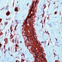
- Formalin-fixed, paraffin-embedded human angiosarcoma stained with VEGF antibody using peroxidase-conjugate and AEC. Note cytoplasmic staining of tumor cells.

- Submitted by
- Invitrogen Antibodies (provider)
- Main image
- Experimental details
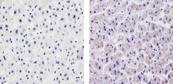
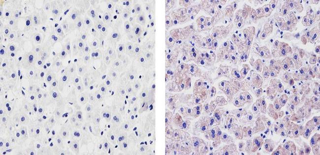
- Immunohistochemistry analysis of VEGF showing staining in the cytoplasm of paraffin-embedded human liver tissue (right) compared to a negative control without primary antibody (left). To expose target proteins, antigen retrieval was performed using 10mM sodium citrate (pH 6.0), microwaved for 8-15 min. Following antigen retrieval, tissues were blocked in 3% H2O2-methanol for 15 min at room temperature, washed with ddH2O and PBS, and then probed with a VEGF Rabbit Polyclonal Antibody (Product # PA5-16754) diluted in 3% BSA-PBS at a dilution of 1:100 for 1 hour at 37ºC in a humidified chamber. Tissues were washed extensively in PBST and detection was performed using an HRP-conjugated secondary antibody followed by colorimetric detection using a DAB kit. Tissues were counterstained with hematoxylin and dehydrated with ethanol and xylene to prep for mounting.
- Submitted by
- Invitrogen Antibodies (provider)
- Main image
- Experimental details
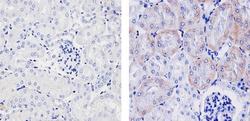
- Immunohistochemistry analysis of VEGF showing staining in the cytoplasm of paraffin-embedded mouse kidney tissue (right) compared to a negative control without primary antibody (left). To expose target proteins, antigen retrieval was performed using 10mM sodium citrate (pH 6.0), microwaved for 8-15 min. Following antigen retrieval, tissues were blocked in 3% H2O2-methanol for 15 min at room temperature, washed with ddH2O and PBS, and then probed with a VEGF Rabbit Polyclonal Antibody (Product # PA5-16754) diluted in 3% BSA-PBS at a dilution of 1:100 for 1 hour at 37ºC in a humidified chamber. Tissues were washed extensively in PBST and detection was performed using an HRP-conjugated secondary antibody followed by colorimetric detection using a DAB kit. Tissues were counterstained with hematoxylin and dehydrated with ethanol and xylene to prep for mounting.

Supportive validation
- Submitted by
- Invitrogen Antibodies (provider)
- Main image
- Experimental details
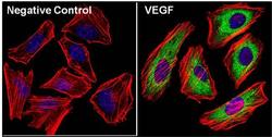
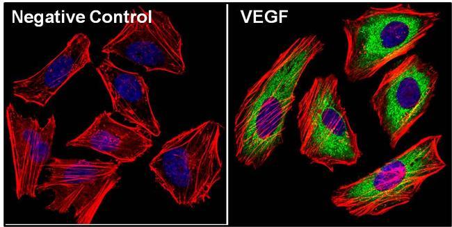
- Immunofluorescent analysis of Vascular Endothelial Growth Factor (VEGF, green) in Hela cells. The cells were fixed with 4% paraformaldehyde, permeabilized with 0.1% Triton X-100 in PBS, and blocked with 3% Blocker BSA (Product # 37525) for 30 minutes at room temperature. Cells were stained with (left panel) or without (right panel) a VEGF polyclonal antibody (Product # PA5-16754) at a dilution of 1:40 for at least 1 hour at room temperature, and then incubated with a Dylight 488 goat anti-rabbit IgG secondary antibody at a dilution of 1:1000 for 45 minutes at room temperature. F-actin (both panels, red) was stained by Dylight 554 Phalloidin (Product # 21834) and nuclei (both panels, blue) were stained with DAPI (Product # 46190). Images were taken at 60X magnification.
- Submitted by
- Invitrogen Antibodies (provider)
- Main image

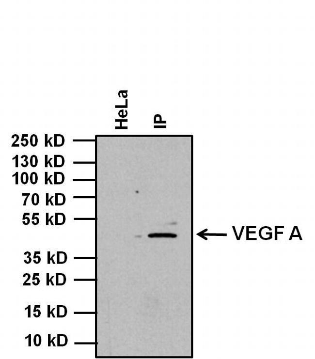
- Experimental details
- Immunoprecipitation of Vascular Endothelial Growth Factor (VEGF) was performed on HeLa cell lysate. Antigen-antibody complexes were formed by incubating 750 µg of whole cell lysate with 3 µg of a VEGF polyclonal antibody (Product # PA5-16754) overnight on a rocking platform at 4C. The immune complexes were captured on 50 µL Protein A/G Plus Agarose (Product # 20423), washed extensively, and eluted with Lane Marker Reducing Sample Buffer (Product # 39000. HeLa cell lysate 20 µg was loaded as a positive control (left lane). Samples were resolved on a 4-20% Tris-Glycing Polyacrylamide gel, transferred to a Nitrocellulose membrane, and blocked with 5% BSA/TBST for at least 1 hour. The membrane was probed with a VEGF polyclonal antibody (Product # PA5-16754) at a concentration of 1 µg/mL in blocking buffer overnight at 4C on a rocking platform, followed by an HRP-conjugated goat anti-rabbit IgG (Fc) secondary antibody (Product # 31463) at a dilution of 1:20,000 for at least 1 hour. Chemiluminescent detection was performed using SuperSignal West Dura (Product # 34076).