Explore
 Validate
 Learn
Learn Western blot
Antibody data
- Antibody Data
- Antigen structure
- References [0]
- Comments [0]
- Validations
- Western blot [2]
- Immunocytochemistry [1]
- Immunohistochemistry [1]
Submit
Validation data
Reference
Comment
Report error
- Product number
- MA5-26823 - Provider product page

- Provider
- Invitrogen Antibodies
- Product name
- DVL2 Monoclonal Antibody (OTI2E11)
- Antibody type
- Monoclonal
- Antigen
- Recombinant protein fragment
- Reactivity
- Human
- Host
- Mouse
- Isotype
- IgG
- Antibody clone number
- OTI2E11
- Vial size
- 100 µL
- Concentration
- 1 mg/mL
- Storage
- -20° C, Avoid Freeze/Thaw Cycles
No comments: Submit comment
Supportive validation
- Submitted by
- Invitrogen Antibodies (provider)
- Main image
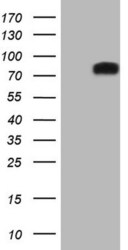
- Experimental details
- Western blot analysis of DVL2 in HEK293T cells in untransfected (Left lane) and transfected (Right lane) samples using 5 µg per lane. The samples were separated by SDS-PAGE and probed with DVL2 (Product # MA5-26823) monoclonal antibody.
- Submitted by
- Invitrogen Antibodies (provider)
- Main image
- Experimental details
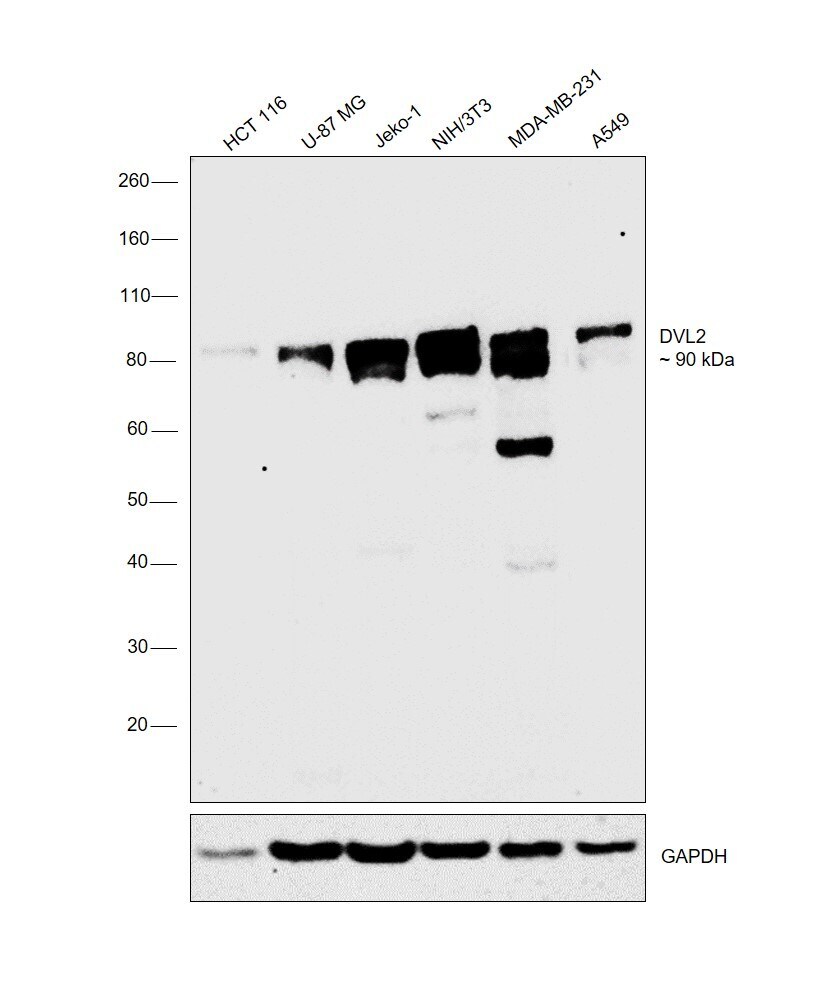
- Western blot was performed using Anti-DVL2 Monoclonal Antibody (OTI2E11) (Product # MA5-26823) and a band at 90 kDa corresponding to DVL2 was observed across the cell lines tested. Whole cell extracts (30 µg lysate) of HCT 116 (Lane 1), U-87 MG (Lane 2), Jeko-1 (Lane 3), NIH/3T3 (Lane 4), MDA-MB-231 (Lane 5) and A549 (Lane 6) were electrophoresed using Novex® NuPAGE® 4-12% % Bis-Tris gel (Product # NP0321BOX). Resolved proteins were then transferred onto a nitrocellulose membrane (Product # IB23001) by iBlot® 2 Dry Blotting System (Product # IB21001). The blot was probed with the primary antibody (1:1000 dilution) and detected by chemiluminescence with Goat anti-Mouse IgG (H+L) Superclonal™ Recombinant Secondary Antibody, HRP (Product # A28177, 1:4000 dilution) using the iBright FL 1000 (Product # A32752). Chemiluminescent detection was performed using Novex® ECL Chemiluminescent Substrate Reagent Kit (Product # WP20005).
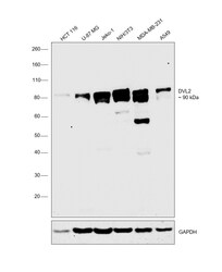
Supportive validation
- Submitted by
- Invitrogen Antibodies (provider)
- Main image
- Experimental details
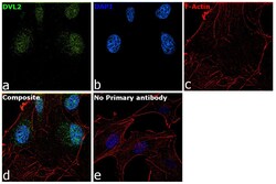
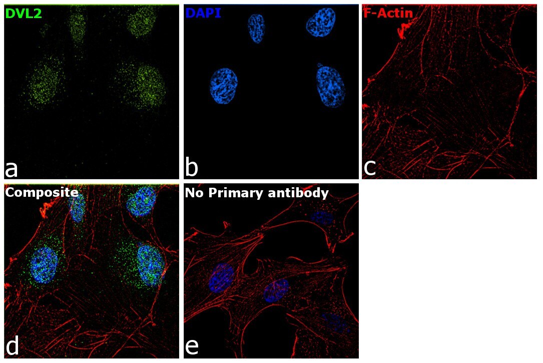
- Immunofluorescence analysis of DVL2 was performed using 70% confluent log phase U-87 MG cells. The cells were fixed with 4% paraformaldehyde for 10 minutes, permeabilized with 0.1% Triton™ X-100 for 15 minutes, and blocked with 2% BSA for 1 hour at room temperature. The cells were labeled with DVL2 Monoclonal Antibody (OTI2E11) (Product # MA5-26823) at 5µg/mL in 0.1% BSA, incubated at 4 degree Celsius overnight and then labeled with Donkey anti-Mouse IgG (H+L) Highly Cross-Adsorbed Secondary Antibody, Alexa Fluor Plus 488 (Product # A32766) at a dilution of 1:2000 for 45 minutes at room temperature (Panel a: green). Nuclei (Panel b: blue) were stained with SlowFade® Gold Antifade Mountant with DAPI (Product # S36938). F-actin (Panel c: red) was stained with Rhodamine Phalloidin (Product # R415, 1:300). Panel d represents the merged image showing nuclear localization. Panel e represents control cells with no primary antibody to assess background. The images were captured at 60X magnification.
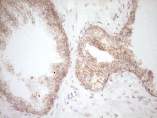
Supportive validation
- Submitted by
- Invitrogen Antibodies (provider)
- Main image
- Experimental details
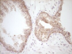
- Immunohistochemistry was performed on paraffin-embedded carcinoma of human prostate tissue. To expose target proteins, 1 mM EDTA in 10mM Tris, pH8.5, 120°C for 3min was used. Following antigen retrieval, tissues were probed with a DVL2 monoclonal antibody (Product # MA5-26823).