Explore
 Validate
 Learn
Learn Western blot
Antibody data
- Antibody Data
- Antigen structure
- References [1]
- Comments [0]
- Validations
- Western blot [5]
- Immunocytochemistry [2]
- Immunohistochemistry [1]
- Chromatin Immunoprecipitation [1]
- Other assay [1]
Submit
Validation data
Reference
Comment
Report error
- Product number
- PA5-78321 - Provider product page

- Provider
- Invitrogen Antibodies
- Product name
- YAP1 Polyclonal Antibody
- Antibody type
- Polyclonal
- Antigen
- Recombinant full-length protein
- Description
- Positive Control: 293T, A431, HeLa, HepG2
- Concentration
- 0.15 mg/mL
Submitted references Interaction of YAP1 and mTOR promotes bladder cancer progression.
Xu M, Gu M, Zhou J, Da J, Wang Z
International journal of oncology 2020 Jan;56(1):232-242
International journal of oncology 2020 Jan;56(1):232-242
No comments: Submit comment
Supportive validation
- Submitted by
- Invitrogen Antibodies (provider)
- Main image
- Experimental details
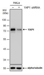
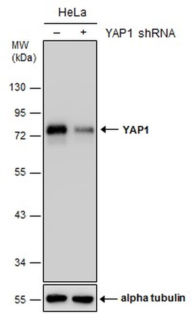
- Western blot analysis of YAP1 in non-transfected (-) and transfected (+) HeLa cells using 30 µg of protein. Samples were separated with 10% SDS-PAGE and incubated with YAP1 polyclonal antibody (Product # PA5-78321) using a dilution of 1:5000 followed by HRP-conjugated anti-rabbit IgG.
- Submitted by
- Invitrogen Antibodies (provider)
- Main image
- Experimental details
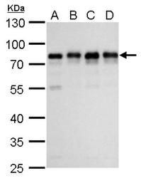
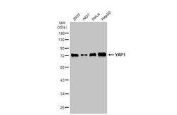
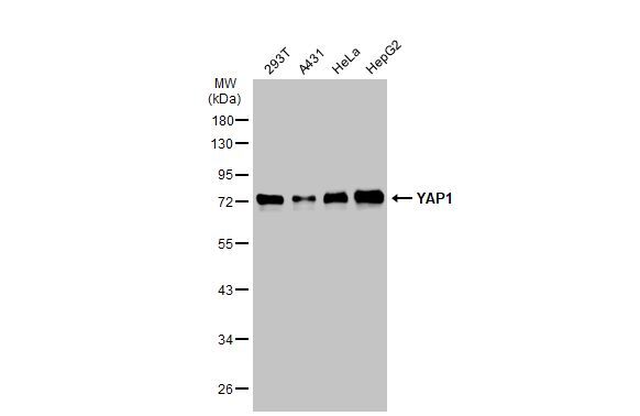
- Western blot analysis of YAP1 in A) 293T whole cell lysate, B) A431 whole cell lysate, C) HeLa whole cell lysate, D) HepG2 whole cell lysate using 30 µg of protein. Samples were separated with 10% SDS-PAGE and incubated with YAP1 polyclonal antibody (Product # PA5-78321) using a dilution of 1:1000.

- Submitted by
- Invitrogen Antibodies (provider)
- Main image

- Experimental details
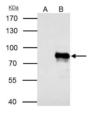
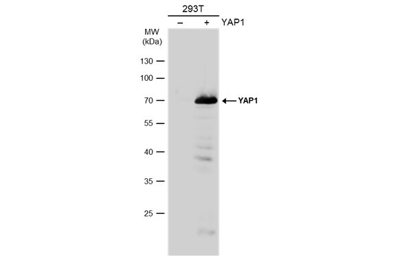
- Western Blot analysis of YAP1 was performed by separating 30 µg of non-transfected (–) and transfected (+) 293T whole cell extracts by 10% SDS-PAGE. Proteins were transferred to a membrane and probed with a YAP1 Polyclonal Antibody (Product # PA5-78321) at a dilution of 1:20000. The HRP-conjugated anti-rabbit IgG antibody was used to detect the primary antibody.
- Submitted by
- Invitrogen Antibodies (provider)
- Main image
- Experimental details
- Western Blot using YAP1 Polyclonal Antibody (Product # PA5-78321). Various whole cell extracts (30 µg) were separated by 10% SDS-PAGE, and the membrane was blotted with YAP1 Polyclonal Antibody (Product # PA5-78321) diluted at 1:1,000. The HRP-conjugated anti-rabbit IgG antibody was used to detect the primary antibody.
- Submitted by
- Invitrogen Antibodies (provider)
- Main image
- Experimental details
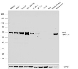
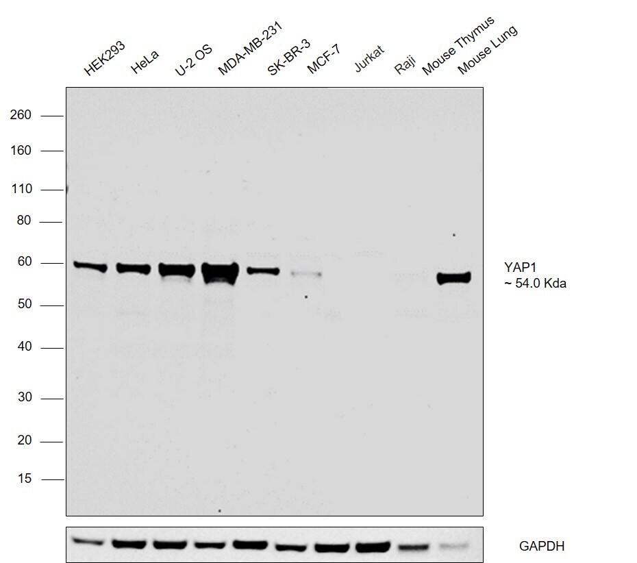
- Western blot analysis was performed on whole cell extracts (30 µg lysate) of HEK293 (Lane 1), HeLa (Lane 2), U-2 OS (Lane 3), MDA-MB-231 (Lane 4), SK-BR-3 (Lane 5), MCF-7 (Lane 6), Jurkat (Lane 7), Raji (Lane 8), Mouse Thymus(Lane 9) and Mouse Lung (Lane 10) and a 54 kDa band corresponding to YAP1 was observed at slightly higher molecular weight across all the cell lines tested except Jurkat and Raji which are reported to be negative. Resolved proteins were then transferred onto a nitrocellulose membrane (Product # IB23001) by iBlot® 2 Dry Blotting System (Product # IB21001).The blot was probed with Anti-YAP1 Polyclonal Antibody (Product # PA5-78321, 1:1000 dilution) and detected by chemiluminescence with Goat anti-Rabbit IgG (H+L) Superclonal™ Recombinant Secondary Antibody, HRP conjugate (Product # A27036, 0.25 µg/ml, 1:4000 dilution) using the iBright FL 1000 (Product # A32752). Chemiluminescent detection was performed using Novex® ECL Chemiluminescent Substrate Reagent Kit (Product # WP20005).
Supportive validation
- Submitted by
- Invitrogen Antibodies (provider)
- Main image
- Experimental details
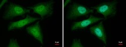
- YAP1 Polyclonal Antibody detects YAP1 protein at cytoplasm and nucleus by immunofluorescent analysis. Sample: HeLa cells were fixed in 4% paraformaldehyde at RT for 15 min. Green: YAP1 protein stained by YAP1 Polyclonal Antibody (Product # PA5-78321) diluted at 1:1,000. Blue: Hoechst 33342 staining.

- Submitted by
- Invitrogen Antibodies (provider)
- Main image

- Experimental details
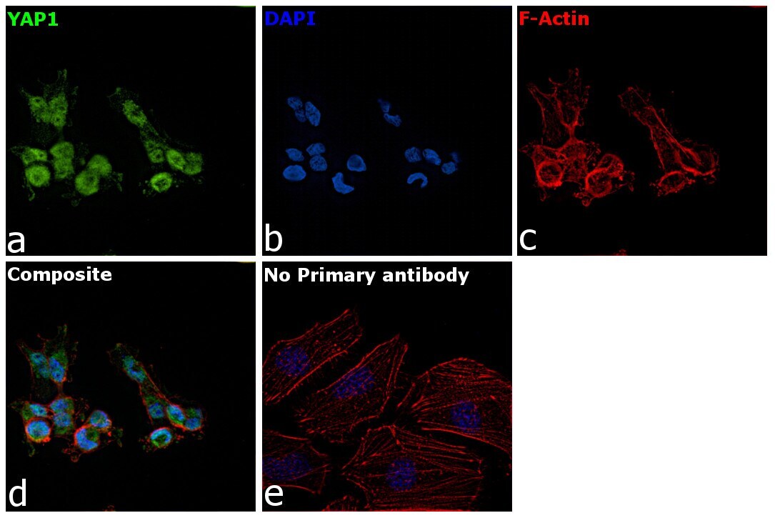
- Immunofluorescence analysis of YAP1 was performed using 70% confluent log phase MDAMB cells. The cells were fixed with 4% paraformaldehyde for 10 minutes, permeabilized with 0.1% Triton™ X-100 for 15 minutes, and blocked with 2% BSA for 1 hour at room temperature. The cells were labeled with YAP1 Polyclonal Antibody (Product # PA5-78321) at 5 microgram/mL in 0.1% BSA, incubated at 4 degree Celsius overnight and then labeled with Goat anti-Rabbit IgG (H+L) Superclonal™ Recombinant Secondary Antibody, Alexa Fluor® 488 conjugate (Product # A27034) at a dilution of 1:2000 for 45 minutes at room temperature (Panel a: green). Nuclei (Panel b: blue) were stained with ProLong™ Diamond Antifade Mountant with DAPI (Product # P36962). F-actin (Panel c: red) was stained with Rhodamine Phalloidin (Product # R415). Panel d represents the merged image showing Nucleus and Cytoplasm localization. Panel e represents control cells with no primary antibody to assess background. The images were captured at 60X magnification.
Supportive validation
- Submitted by
- Invitrogen Antibodies (provider)
- Main image
- Experimental details
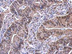
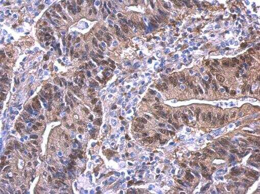
- YAP1 Polyclonal Antibody detects YAP1 protein at nucleus on human colon carcinoma by immunohistochemical analysis. Sample: Paraffin-embedded colon carcinoma. YAP1 Polyclonal Antibody (Product # PA5-78321) dilution: 1:500. Antigen Retrieval: EDTA based buffer, pH 8.0, 15 min.
Supportive validation
- Submitted by
- Invitrogen Antibodies (provider)
- Main image

- Experimental details
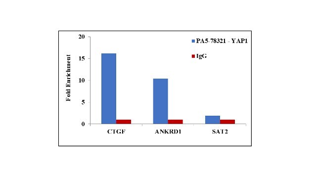
- Chromatin Immunoprecipitation (ChIP) assay of endogenous YAP1 protein using Anti-YAP1 Antibody: ChIP was performed using Anti-YAP1 Polyclonal Antibody (Product # PA5-78321, 2.5 µg) on sheared chromatin from MDA-MB-231 cells using the MAGnify ChIP System kit (Product # 49-2024). Normal Rabbit IgG was used as a negative IP control. The purified DNA was analyzed by qPCR using primers binding to ANKRD1 and CTGF transcriptional start site and SAT2 satellite repeats. Data is presented as fold enrichment of the antibody signal versus the negative control IgG using the comparative CT method.
Supportive validation
- Submitted by
- Invitrogen Antibodies (provider)
- Main image
- Experimental details
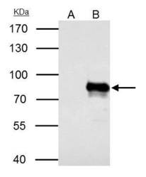
- YAP1 Polyclonal Antibody immunoprecipitates YAP1 protein in IP experiments. IP samples: HeLa whole cell extract. A. Control with 4 µg of preimmune Rabbit IgG. B. Immunoprecipitation of YAP1 protein by 4 µg YAP1 Polyclonal Antibody (Product # PA5-78321). 7.5 % SDS-PAGE. The immunoprecipitated YAP1 protein was detected by YAP1 Polyclonal Antibody (Product # PA5-78321) diluted at 1:500.