Explore
 Validate
 Learn
Learn Western blot
 Immunoprecipitation
Antibody data
- Antibody Data
- Antigen structure
- References [4]
- Comments [0]
- Validations
- Western blot [1]
- Immunohistochemistry [3]
Submit
Validation data
Reference
Comment
Report error
- Product number
- NB100-56126 - Provider product page

- Provider
- Novus Biologicals
- Proper citation
- Novus Cat#NB100-56126, RRID:AB_2069213
- Product name
- Rabbit Polyclonal Caspase-14 Antibody
- Antibody type
- Polyclonal
- Description
- Unpurified. This polyclonal antibody recognizes the proform of caspase-14 (~-28-32 kDa), and the large (~14-21 kDa) and small (~10-11 kDa) of active/cleaved caspase-14.
- Reactivity
- Human, Mouse, Rat, Canine
- Host
- Rabbit
- Isotype
- IgG
- Vial size
- 0.05 ml
- Storage
- Store at 4C short term. Aliquot and store at -20C long term. Avoid freeze-thaw cycles.
Submitted references Dual Inhibition of PI3K/Akt and mTOR by the Dietary Antioxidant, Delphinidin, Ameliorates Psoriatic Features In Vitro and in an Imiquimod-Induced Psoriasis-Like Disease in Mice.
Association of caspase-14 and filaggrin expression with keratinization of the oral mucosa and reconstruction culture rat models.
Tumor-associated alterations in caspase-14 expression in epithelial malignancies.
Early processing of Bid and caspase-6, -8, -10, -14 in the canine brain during cardiac arrest and resuscitation.
Chamcheu JC, Adhami VM, Esnault S, Sechi M, Siddiqui IA, Satyshur KA, Syed DN, Dodwad SM, Chaves-Rodriquez MI, Longley BJ, Wood GS, Mukhtar H
Antioxidants & redox signaling 2017 Jan 10;26(2):49-69
Antioxidants & redox signaling 2017 Jan 10;26(2):49-69
Association of caspase-14 and filaggrin expression with keratinization of the oral mucosa and reconstruction culture rat models.
Murakami H, Okamura K, Aoki S, Sakagami R, Yamazaki J
Journal of periodontal research 2014 Dec;49(6):703-10
Journal of periodontal research 2014 Dec;49(6):703-10
Tumor-associated alterations in caspase-14 expression in epithelial malignancies.
Krajewska M, Kim H, Shin E, Kennedy S, Duffy MJ, Wong YF, Marr D, Mikolajczyk J, Shabaik A, Meinhold-Heerlein I, Huang X, Banares S, Hedayat H, Reed JC, Krajewski S
Clinical cancer research : an official journal of the American Association for Cancer Research 2005 Aug 1;11(15):5462-71
Clinical cancer research : an official journal of the American Association for Cancer Research 2005 Aug 1;11(15):5462-71
Early processing of Bid and caspase-6, -8, -10, -14 in the canine brain during cardiac arrest and resuscitation.
Krajewska M, Rosenthal RE, Mikolajczyk J, Stennicke HR, Wiesenthal T, Mai J, Naito M, Salvesen GS, Reed JC, Fiskum G, Krajewski S
Experimental neurology 2004 Oct;189(2):261-79
Experimental neurology 2004 Oct;189(2):261-79
No comments: Submit comment
Supportive validation
- Submitted by
- Novus Biologicals (provider)
- Main image
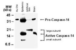
- Experimental details
- Western Blot: Caspase-14 Antibody [NB100-56126] - Analysis of Caspase-14. Tissue lysates (50 ug/lane) and recombinant human Caspase-14 were (Hu C14, 15 ng) were western blotted with Caspase-14 antibody at 1:2000. The antibody detected both the proform of caspase-14, and the large and small subunits of active/cleaved caspase-14.

Supportive validation
- Submitted by
- Novus Biologicals (provider)
- Main image
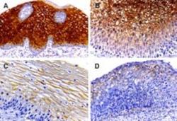
- Experimental details
- Immunohistochemistry-Paraffin: Caspase-14 Antibody [NB100-56126] - Tissue sections of human cervix stained using this antibody at 1:2000. A. Normal cervix (squamous epithelium). B. CIN1 (low-grade squamous intraepithelial lesion, mild dysplasia). C. CIN2 (high-grade squamous intraepithelial lesion, moderate dysplasia. D. CIN3 (high-grade squamous intraepithelial lesion; severe dysplasia-carcinoma in situ. In normal cervi, caspase-14 staining was found most in the midzone layer, but was absent from the basal/parabasal cell layer where mitotically active cells are known to reside. This suggests induction of caspase-14 expression with differentiation. Caspase-14 expression declined progressively during malignant transformation as the histologic severity of the cervical atypia advanced from CIN1 to CIN3. Hematoxylin-eosin counterstain.

- Submitted by
- Novus Biologicals (provider)
- Main image
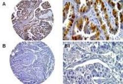
- Experimental details
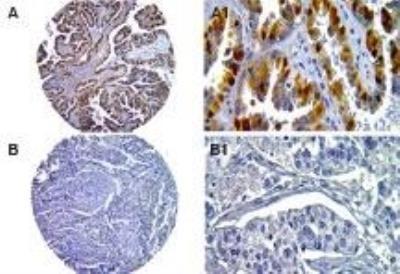
- Immunohistochemistry-Paraffin: Caspase-14 Antibody [NB100-56126] - Human ovarian cancer tissue microarray stained for Caspase-14 expression using this antibody at 1:2000. Low (A) and high (B) stage ovarian tumor tissue cores. High magnification from areas of the tissue cores (A1 and B1). Decreased Caspase-14 expression was seen in the high grade, compared to the low grade tumor. Hematoxylin-eosin counterstain.
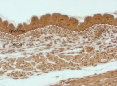
- Submitted by
- Novus Biologicals (provider)
- Main image
- Experimental details
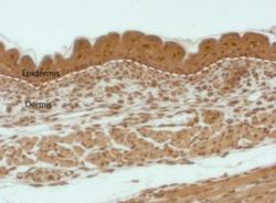
- Immunohistochemistry: Caspase-14 Antibody [NB100-56126] - Tissue sections of mouse skin at E17 stained using this antibody at 1:500.