Explore
 Validate
 Learn
Learn Western blot
Antibody data
- Antibody Data
- Antigen structure
- References [2]
- Comments [0]
- Validations
- Western blot [4]
- Immunohistochemistry [2]
- Flow cytometry [4]
Submit
Validation data
Reference
Comment
Report error
- Product number
- PA1-16857 - Provider product page

- Provider
- Invitrogen Antibodies
- Product name
- Beclin 1 Polyclonal Antibody
- Antibody type
- Polyclonal
- Antigen
- Synthetic peptide
- Description
- The target sequence has 88% sequence homology with rat.
- Concentration
- 1 mg/mL
Submitted references Reprogramming of Amino Acid Transporters to Support Aspartate and Glutamate Dependency Sustains Endocrine Resistance in Breast Cancer.
Intracardiac administration of ephrinA1-Fc preserves mitochondrial bioenergetics during acute ischemia/reperfusion injury.
Bacci M, Lorito N, Ippolito L, Ramazzotti M, Luti S, Romagnoli S, Parri M, Bianchini F, Cappellesso F, Virga F, Gao Q, Simões BM, Marangoni E, Martin LA, Comito G, Ferracin M, Giannoni E, Mazzone M, Chiarugi P, Morandi A
Cell reports 2019 Jul 2;28(1):104-118.e8
Cell reports 2019 Jul 2;28(1):104-118.e8
Intracardiac administration of ephrinA1-Fc preserves mitochondrial bioenergetics during acute ischemia/reperfusion injury.
Torres MJ, McLaughlin KL, Renegar RH, Valsaraj S, Whitehurst KS, Sharaf OM, Sharma UM, Horton JL, Sarathy B, Parks JC, Brault JJ, Fisher-Wellman KH, Neufer PD, Virag JAI
Life sciences 2019 Dec 15;239:117053
Life sciences 2019 Dec 15;239:117053
No comments: Submit comment
Supportive validation
- Submitted by
- Invitrogen Antibodies (provider)
- Main image

- Experimental details
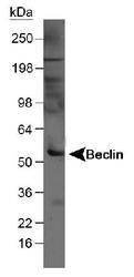
- Western Blot detection of Beclin in mouse liver tissue lysate (50 µg) using Product # PA1-16857. ECL Western Blot detection at 1 minute.
- Submitted by
- Invitrogen Antibodies (provider)
- Main image
- Experimental details
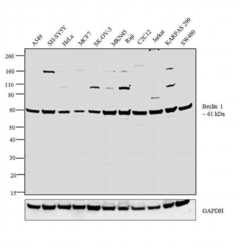
- Western blot analysis was performed on whole cell extracts (30 µg lysate) of A549 (Lane 1), SH-SY5Y (Lane 2), HeLa (Lane 3), MCF7 (Lane 4), SK-OV-3 (Lane 5), MKN45 (Lane 6), Raji (Lane 7), C2C12 (Lane 8), Jurkat (Lane 9), KARPAS 299 (Lane 10) and SW480 (Lane 11). The blot was probed with Anti-Beclin 1 Polyclonal Antibody (Product # PA1-16857, 1:500 dilution) and detected by chemiluminescence using Goat anti-Rabbit IgG (H+L) Superclonal™ Secondary Antibody, HRP conjugate (Product # A27036, 0.25 µg/mL, 1:4000 dilution). A 61 kDa band corresponding to Beclin 1 was observed across the cell lines tested.

- Submitted by
- Invitrogen Antibodies (provider)
- Main image
- Experimental details
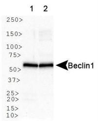
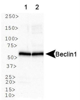
- Western blot analysis of Beclin 1 in Lane 1: human brain. Lane 2: mouse brain. Samples were incubated in Beclin 1 polyclonal antibody (Product # PA1-16857).
- Submitted by
- Invitrogen Antibodies (provider)
- Main image

- Experimental details
- Western blot analysis of Beclin 1 in 1.0 mg/mL HeLa lysate. Samples were incubated in Beclin 1 polyclonal antibody (Product # PA1-16857). This experiment was performed under reducing conditions using the 12-230 kDa separation system.

Supportive validation
- Submitted by
- Invitrogen Antibodies (provider)
- Main image

- Experimental details
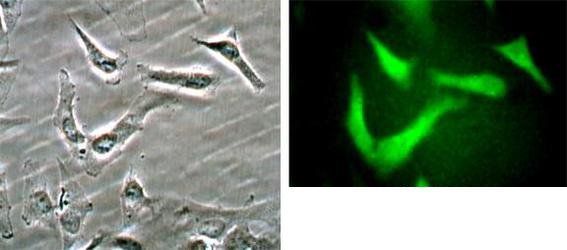
- Beclin 1 staining in Hela cells detected with a Dylight 488 labeled secondary antibody using Product # PA1-16857.
- Submitted by
- Invitrogen Antibodies (provider)
- Main image
- Experimental details
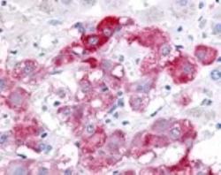
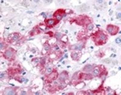
- Immunohistochemical analysis of Beclin 1 in Pheochromocytes of the Adrenal Medulla. Samples were incubated in Beclin 1 polyclonal antibody (Product # PA1-16857). Antibody shown in red. Magnification 40X.
Supportive validation
- Submitted by
- Invitrogen Antibodies (provider)
- Main image
- Experimental details
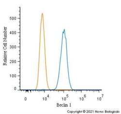
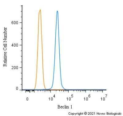
- Flow cytometry of Beclin 1 in Neuro2a cells (blue) and a matched isotype control (orange). Samples were incubated in Beclin 1 polyclonal antibody (Product # PA1-16857) using a dilution of 1.0 µg/mL for 30 minutes at room temperature followed by a Rabbit IgG (H+L) Cross-Adsorbed Secondary Antibody, Dylight™ 550 (Product # SA5-10033). Cells were fixed with 4% PFA and then permeabilized with 0.1% saponin.
- Submitted by
- Invitrogen Antibodies (provider)
- Main image
- Experimental details
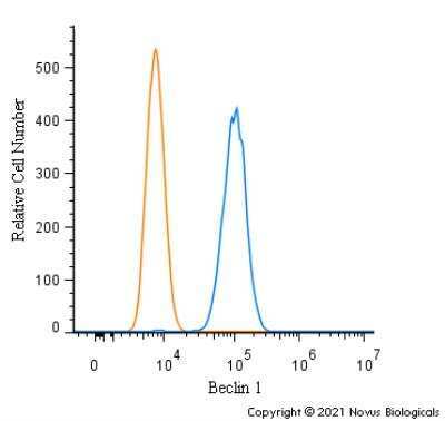
- Flow cytometry of Beclin 1 in HepG2 cells (blue) and a matched isotype control (orange). Samples were incubated in Beclin 1 polyclonal antibody (Product # PA1-16857) using a dilution of 1.0 µg/mL for 30 minutes at room temperature followed by a Rabbit IgG (H+L) Cross-Adsorbed Secondary Antibody, Dylight™ 550 (Product # SA5-10033). Cells were fixed with 4% PFA and then permeabilized with 0.1% saponin.
- Submitted by
- Invitrogen Antibodies (provider)
- Main image

- Experimental details
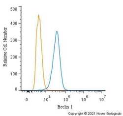
- Flow cytometry of Beclin 1 in THP-1 cells (blue) and a matched isotype control (orange). Samples were incubated in Beclin 1 polyclonal antibody (Product # PA1-16857) using a dilution of 1.0 µg/mL for 30 minutes at room temperature followed by a Rabbit IgG (H+L) Cross-Adsorbed Secondary Antibody, Dylight™ 550 (Product # SA5-10033). Cells were fixed with 4% PFA and then permeabilized with 0.1% saponin.
- Submitted by
- Invitrogen Antibodies (provider)
- Main image
- Experimental details
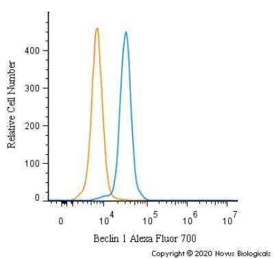
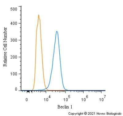
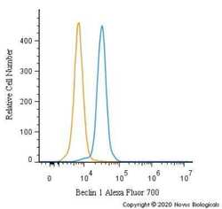
- Flow cytometry of Beclin 1 in HeLa cells. Samples were incubated in Beclin 1 polyclonal antibody (Product # PA1-16857) using a dilution of 5 µg/mL for 30 minutes at room temperature. Antibody (blue) and a matched isotype control (orange). Cells were fixed with 4% PFA and then permeabilized with 0.1% saponin. Both antibodies were conjugated to Alexa Fluor 700.