 Explore
 Validate
 Learn
Learn Western blot
 ELISA
ELISAAntibody data
- Antibody Data
- Antigen structure
- References [2]
- Comments [0]
- Validations
- Western blot [1]
- Immunocytochemistry [1]
- Blocking/Neutralizing [1]
Submit
Validation data
Reference
Comment
Report error
- Product number
- AF-202-NA - Provider product page

- Provider
- R&D Systems
- Product name
- Human IL-2 Antibody
- Antibody type
- Polyclonal
- Description
- Antigen Affinity-purified. Detects human IL-2 in direct ELISAs and Western blots. In direct ELISAs, greater than 50% cross-reactivity with recombinant rabbit IL-2 is observed and less than 20% cross-reactivity with recombinant bovine IL-2, recombinant mouse IL-2 and recombinant rat IL-2 is observed.
- Reactivity
- Human
- Host
- Goat
- Conjugate
- Unconjugated
- Antigen sequence
P60568- Isotype
- IgG
- Vial size
- 100 ug
- Concentration
- LYOPH
- Storage
- Use a manual defrost freezer and avoid repeated freeze-thaw cycles. 12 months from date of receipt, -20 to -70 °C as supplied. 1 month, 2 to 8 °C under sterile conditions after reconstitution. 6 months, -20 to -70 °C under sterile conditions after reconstitution.
Submitted references Natural and induced CD4+CD25+ cells educate CD4+CD25- cells to develop suppressive activity: the role of IL-2, TGF-beta, and IL-10.
Natural and induced CD4+CD25+ cells educate CD4+CD25- cells to develop suppressive activity: the role of IL-2, TGF-beta, and IL-10.
Zheng SG, Wang JH, Gray JD, Soucier H, Horwitz DA
Journal of immunology (Baltimore, Md. : 1950) 2004 May 1;172(9):5213-21
Journal of immunology (Baltimore, Md. : 1950) 2004 May 1;172(9):5213-21
No comments: Submit comment
Supportive validation
- Submitted by
- R&D Systems (provider)
- Main image

- Experimental details
- Detection of Human IL-2 by Western Blot. Western blot shows lysates of monensin treated human peripheral blood mononuclear cells (PBMCs) with no additional treatment (-) or additionally treated (+) with 0.5ug/mL calcium ionomycin (Iono) and 50ng/mL PMA overnight. PVDF membrane was probed with 0.5 µg/mL of Goat Anti-Human IL-2 Antigen Affinity-purified Polyclonal Antibody (Catalog # AF-202-NA) followed by HRP-conjugated Anti-Goat IgG Secondary Antibody (Catalog # HAF017). A specific band was detected for IL-2 at approximately 14 kDa (as indicated). This experiment was conducted under reducing conditions and using Immunoblot Buffer Group 1.

Supportive validation
- Submitted by
- R&D Systems (provider)
- Main image

- Experimental details
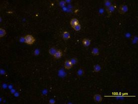
- IL-2 in Human PBMCs. IL-2 was detected in immersion fixed human peripheral blood mononuclear cells (PBMCs) stimulated with PMA, ionomyocin, and monensin using Goat Anti-Human IL-2 Antigen Affinity-purified Polyclonal Antibody (Catalog # AF-202-NA) at 10 µg/mL for 3 hours at room temperature. Cells were stained using the NorthernLights™ 557-conjugated Anti-Goat IgG Secondary Antibody (yellow; Catalog # NL001) and counter-stained with DAPI (blue). View our protocol for Fluorescent ICC Staining of Non-adherent Cells.
Supportive validation
- Submitted by
- R&D Systems (provider)
- Main image

- Experimental details
- Cell Proliferation Induced by IL-2 and Neutralization by Human IL-2 Antibody. Recombinant Human IL-2 (Catalog # 202-IL) stimulates proliferation in the CTLL-2 mouse cytotoxic T cell line in a dose-dependent manner (orange line) as measured by Resazurin (Catalog # AR002). Proliferation elicited by Recombinant Human IL-2 (2 ng/mL) is neutralized (green line) by increasing concentrations of Goat Anti-Human IL-2 Antigen Affinity-purified Polyclonal Antibody (Catalog # AF-202-NA). The ND50 is typically
